- Introduction to the digestive system—structure and four layers of the alimentary canal
- Mouth, pharynx, esophagus and stomach – structure and function
- Small intestine, large intestine, and rectum—structure and function
- Digestive glands — salivary glands, liver, pancreas
- Stepwise digestion of carbohydrates, proteins and fats
- Hormones and disorders of the absorption, assimilation and digestive systems
- Respiratory system—the journey from the nostrils to the lungs
- Lungs, pleura and lung volume capacities
- Respiratory mechanisms — inhalation and exhalation
- Exchange and transport of gases (O₂ and CO₂)
- Regulation of respiration and diseases of the respiratory system
- Circulatory system – blood composition, blood groups and coagulation
- Structure of the heart, cardiac cycle and motor system
- Blood vessels and types of circulation
- Diseases of the blood pressure, lymphatic system, and circulatory system
- Excretory system – nitrogenous wastes and the structure of the kidneys
- The three-step process of nephron and urine formation
- Hormonal regulation, dialysis and diseases of the excretory system
Imagine four massive factories running simultaneously inside your body, nonstop, nonstop, 24 hours a day—one factory breaks down food and converts it into energy (the digestive system), another draws oxygen from the air and delivers it to the blood (the respiratory system), a third runs a pumping station that beats nearly a million times a day, delivering every drop of blood to every corner of the body (the circulatory system), and a fourth filters out toxic wastes and expels them (the excretory system). The most amazing thing is—you don't even have to press a button to start any of these factories! Whether you're sleeping, reading, or playing badminton, these four systems work automatically, nonstop. This is the true engineering of the human body—what scientists call "human physiology." Let's take a peek inside these four amazing systems to see what your body is doing for you every moment.
1 Introduction to the Digestive System - Structure of the Alimentary Canal
From the moment you put a morsel of bread in your mouth, until the energy from it reaches your muscles – a long, highly organized journey begins. This journey takes place in your Alimentary Canal, which is a continuous, muscular, open tube about 8-10 meters long, starting from the mouth and ending at the anus – that is, a tunnel 5-6 times longer than your height, which coils and ends in your stomach! The function of the digestive system is to convert the complex nutrients and large molecules of food into simple, small and soluble substances with the help of various chemical reactions and enzymes – this process is called digestion.
The digestive system consists of two types of organs—first, the organs of the alimentary canal (mouth, pharynx, esophagus, stomach, small intestine, large intestine, anus) through which food passes directly; second, the digestive glands (salivary glands, liver, pancreas) which release digestive juices into the alimentary canal through ducts. Digestion occurs primarily by hydrolysis—in which H+ and OH– ions of water bind to complex molecules (such as starch, protein, fat) and break them down into simpler molecules; enzymes act only as catalysts in this process; they are not destroyed themselves.
Four layers of the gut wall
The alimentary canal is not a simple hollow tube, but a highly organized, multilayered structure. From the inside out, there are four layers:
- Mucosa – The innermost layer, composed of simple columnar epithelium; it contains goblet cells that secrete mucus to lubricate and protect the wall.
- Submucosa – It contains blood vessels, lymph vessels and crypts of Lieberkuhn.
- Muscularis – Made up of two layers of circular muscle fibers and longitudinal muscle fibers; this layer produces peristalsis and propels food forward.
- Serosa – The outermost protective covering layer.
Peristalsis is such a powerful movement that even if you swallow food while lying face down or standing on your head, the food will still go against gravity and reach the stomach – because it depends not on gravity but on the undulating motion of the muscular wall.
Three Major Functions of the Digestive System
- Digestion – breaking down complex molecules into simpler ones
- Absorption of digested food – transporting simple nutrients into the blood/lymph
- Propulsion – Propulsion of food from the mouth to the anus
◆ alimentary canal ◆ Hydrolysis ◆ mucosal layer ◆ muscular layer ◆ Peristalsis ◆ Goblet cells
2 Mouth, pharynx, esophagus and stomach – structure and function
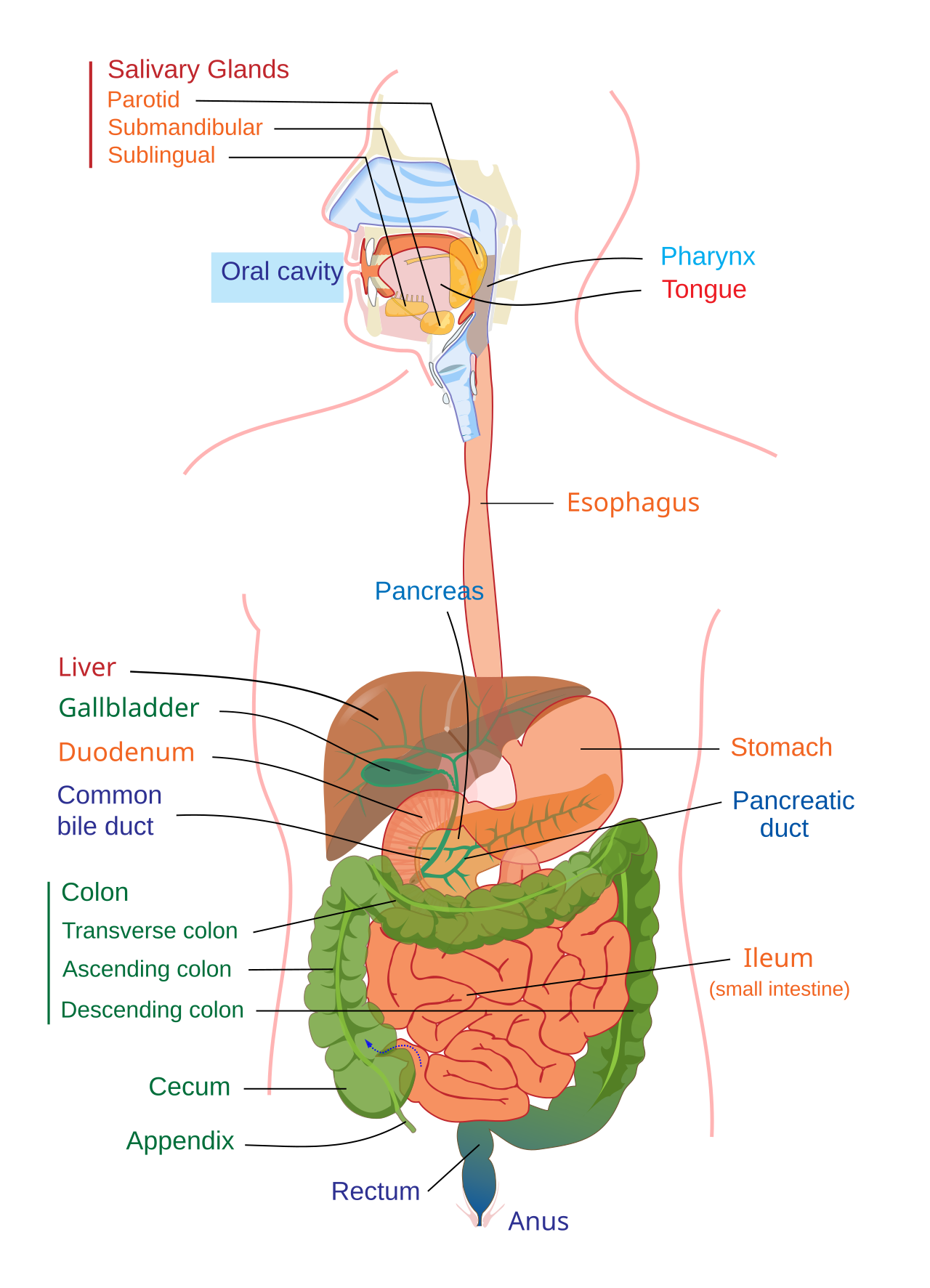
🖼️ Human digestive system – complete structure of the alimentary canal and attached glands from the mouth to the anus
Mouth
The first entrance to the digestive system is the mouth—a bowl-shaped organ with the hard palate above and the soft palate below. The tongue is connected to the floor of the mouth by the frenulum linguae. Taste buds on the tongue are arranged regionally—the front part of the tongue is most sensitive to sweet tastes, the front edge to salty tastes, the middle edge to sour tastes, and the back part to bitter tastes.
Teeth
The teeth in each jaw are located in a mould of the gums, a system called thecodont. In humans, two sets of teeth develop during a lifetime—hence the diphyodont system: first the milk teeth (20 in total), then the permanent teeth (32 in total).
| Type | Number (per jaw) | Work | exit age |
|---|---|---|---|
| Incisors | 4 | nibble-bite | 6–8 months |
| Canines | 2 | tear apart | 16–20 months |
| Premolars | 4 | to chew | 10–11 years |
| Molars | 6 | grinding and chewing | 6-7 years, 12-13 years, 17-25 years (wisdom) |
Enamel, the layer covering teeth, is the hardest substance in the human body—harder than bone! It's composed primarily of calcium phosphate (hydroxyapatite). However, excessive sugar and acid can slowly erode it—the root cause of tooth decay.
Pharynx
A flask-shaped structure located behind the mouth that serves as a common passage for both the respiratory and digestive systems. It has three parts: the nasopharynx (air passage only), the oropharynx (air and food passage), and the laryngopharynx (laryngopharynx).
Oesophagus
A narrow muscular tube about 25 cm long, which passes through the neck and chest, crossing the diaphragm and enters the abdominal cavity. Its mucous glands keep the passage lubricated and juicy, so that the bolus of food easily reaches the stomach through peristaltic movement. While swallowing, the epiglottis (gill lid) momentarily closes the entrance to the trachea, so that food does not accidentally go into the windpipe – this is the reason why our breathing stops momentarily while swallowing.
💡 Epiglottis — the body's automatic switch: Every time you swallow, a small cartilaginous flap called the epiglottis covers the larynx to prevent food from going down the wrong tube. This process happens thousands of times throughout the day, unconsciously, and correctly—and if we accidentally swallow and breathe at the same time, we choke, which shows how intricately this switching mechanism is designed.
Stomach
A muscular, J-shaped structure located between the esophagus and duodenum, behind the diaphragm in the left part of the abdominal cavity. It is a flexible organ that can hold 1 to 3 liters of food. The stomach has three parts:
- Cardiac Portion – The left large portion, where the esophagus enters.
- Fundus Portion – Located just below the diaphragm.
- Pyloric Portion – The right small portion where the small intestine joins.
The stomach wall contains two types of glands—the gastric gland, in which zymogen cells secrete pepsinogen and oxyntic/parietal cells secrete hydrochloric acid (HCl); and the pyloric gland, which secretes mucus. Gastric juice is a highly acidic fluid (pH 1-2) consisting of water (98%), HCl (about 0.5%), mucous mucin, and two enzymes—pepsin and lipase.
How does the stomach not digest itself?The stomach's gastric juice is so acidic (pH 1-2) that it can dissolve even metal—yet it doesn't digest itself because it's protected by a protective layer of mucus. When this layer weakens, an ulcer forms.
The stomach contains two sphincters—the cardiac sphincter, which prevents acidic food from flowing back into the esophagus; and the pyloric sphincter, which divides the stomach and small intestine. Food typically remains in the stomach for 3-4 hours, where pepsin and HCl combine to partially digest proteins.
◆ Thecodont ◆ Diphyodont ◆ Epiglottis ◆ gastric juice ◆ Pepsinogen ◆ oxyntic cells
3 Small intestine, large intestine and rectum – structure and function
Small Intestine
The tube, approximately 7 meters long and 2.5 cm wide, begins at the pyloric portion of the stomach and ends at the large intestine—where most digestion and absorption occurs. It has three parts:
- Duodenum—The smallest C-shaped portion, connected to the stomach; bile from the liver and pancreatic juice from the pancreas meet here (the sphincter of Oddi is located at its opening). Brunner's glands are found in its wall, which secrete an alkaline fluid and protect the duodenum from acidic gastric juice.
- Jejunum—middle portion, about 2 meters long; here the main absorption occurs by enterocytes.
- Ileum—The last and longest part (about 4 meters), coiled; it absorbs bile salts and vitamin B12, which are not absorbed in the jejunum.
microscopic structure of the intestinal wall
The mucosa and submucosa fold to form a Kerckring ring, on which finger-like villi are found. At the base of the villi are the crypts of Lieberkuhn, which contain goblet cells (secreting mucus), intestinal cells (secreting water and enzymes), and Paneth cells (secreting the bactericidal lysozyme enzyme). Each villus contains a central lacteal and a network of blood capillaries. Microvilli, which are extensions of the plasma membrane, are also found on the epithelial cells of the villi.
💡 Textile Engineering Miracle of the Small Intestine: The villi and their microvilli increase the internal absorptive surface of the small intestine by approximately 1000 times. If the inner surface of the small intestine were laid out flat, it would cover an area approximately the size of a tennis court—a classic example of the body's engineering efficiency.
Large Intestine
A tube approximately 1.5 meters long, connected to the small intestine, where specialized bacteria help break down undigested food through fermentation. Its main functions are the absorption of water and minerals and the excretion of undigested food. It has three main parts:
- Caecum—connected to the small intestine; from its first part arises the vermiform appendix, a vestigial organ in humans but large and functional in herbivorous mammals.
- Colon – Inverted 'U' shaped part, divided into four subdivisions – ascending, transverse, descending and sigma-shaped (S-shaped) colon.
- Rectum—The final part of the alimentary canal; its last 3 cm is called the anal canal, which opens into the anus. Here, two types of sphincters, internal and external, control bowel movements.
In humans, the appendix is a useless/vestigial organ, but in herbivorous animals like rabbits, it is an active and vital organ for digesting cellulose. This is a prime example of the traces of evolution in the human body.
Egestion/Defaecation: Undigested parts (plant fibers) and unabsorbed digested matter are temporarily stored in the rectum, where increased water absorption forms semisolid stool. The defecation reflex relaxes the sphincter muscle and expels the stool—usually once or twice every 24 hours.
◆ Duodenum ◆ Villi ◆ Microvilli ◆ Lacteal ◆ appendix ◆ bowel movement reflex
4 Digestive glands — salivary glands, liver, pancreas
Salivary Gland
Saliva is a mixture of serous fluid and viscous mucus—the liquid portion moistens food, while the mucous portion acts as a lubricant. Humans secrete approximately 1,000 to 1,200 milliliters of saliva daily—that's nearly a full water bottle full of saliva, every day!
There are three pairs of salivary glands:
- Parotid gland—largest, located anterior to the ear; secretes primarily aqueous, amylase-rich saliva.
- Sub-mandibular gland – Mixed gland (both water and mucus), at the angle of the lower jaw.
- Sublingual gland – Smallest gland under the tongue, secreting mainly mucus.
Functions of saliva: Partial digestion of starch (conversion into maltose by amylase/ptyalin enzymes), making food smooth and swallowable (bolus formation), cleaning of teeth, mouth and tongue, destruction of tooth decay causing bacteria by lysozyme, and stimulating the taste buds by dissolving food particles.
It's an interesting empirical fact—saliva secretion is controlled not just by the taste of food, but also by the mechanical act of chewing. Seeing, smelling, or even thinking or talking about food can also trigger salivation—this is due to stimulation of nerve impulses from the brain.
Liver
The largest and most important digestive gland in the human body, a triangular organ located in the upper right part of the abdomen, below the diaphragm. It is composed of approximately 100,000 tiny hexagonal structural-functional units—liver lobules—that is, a million mini chemical plants operating within the body!
Major functions of the liver:
- Bile formation – helps emulsify and digest fats
- Metabolism – the main center for the metabolism of carbohydrates, fats, proteins, and amino acids
- Glycogen storage—storing glucose as glycogen and regulating blood sugar
- Urea production – conversion of toxic ammonia into urea
- Plasma protein synthesis – prothrombin, fibrinogen (for blood clotting) and heparin
- Vitamin and mineral stores – vitamins A, D, E, K, B12 and minerals such as iron and copper
- Blood purification – Kupffer cells break down dead RBCs and hemoglobin.
- Detoxification – eliminating the effects of toxins and drugs in the blood
The liver has a tremendous regeneration capacity—it can regrow even if a large portion of its tissue is removed. Bile contains no digestive enzymes, yet it is essential for fat digestion—it is a yellow-green, alkaline fluid (pH approximately 8), whose main components are bile salts (sodium glycocholate and sodium taurocholate) and bile pigments—bilirubin and biliverdin. Bile is stored and concentrated in the gallbladder and released into the duodenum in response to food intake.
How is a substance without enzymes so important?Bile contains no enzymes, yet it's the heart of fat digestion—because it breaks down large globules of fat into smaller droplets through emulsification, much like dish soap breaks down oil. This increases the surface area for enzymes, allowing them to work faster.
Pancreas
It is a mixed gland—endocrinely, it regulates blood sugar by producing insulin and glucagon hormones (from the islets of Langerhans); exocrinely, it produces pancreatic juice, which helps in the digestion of proteins, fats, and carbohydrates in the intestines. It is a 6-8 inch long, U-shaped gland, surrounded by the liver, duodenum, and spleen. Its secretions reach the duodenum through the pancreatic duct.
◆ Parotid gland ◆ Ptyalin/Amylase ◆ hepatic lobes ◆ bile salts ◆ bilirubin ◆ Islets of Langerhans
5 Stepwise digestion of carbohydrates, proteins and fats
The three main nutrients in food—carbohydrates, proteins, and fats—are broken down step by step by different enzymes in different parts of the digestive tract until they are reduced to their simplest absorbable units. The general sequence of the entire process is: Ingestion → Digestion → Absorption → Assimilation → Egestion.
(A) Digestion of carbohydrates
| place | Enzymes/Sources | verb |
|---|---|---|
| buccal cavity | Salivary amylase (ptyalin) | Cooked starch → maltose (sweet-tasting disaccharide); raw starch → dextrin |
| stomach | — | Salivary amylase continues to act until the medium is acidified by HCl; the main digestion does not occur here. |
| duodenum | pancreatic amylase | Starch, glycogen → maltose + oligosaccharides; digests all starch in about half an hour |
| small intestine | Maltase, sucrase, lactase | Maltose → 2 glucose; sucrose → glucose + fructose; lactose → glucose + galactose |
Final absorbable form: Monosaccharides — glucose, fructose, galactose.
(B) digestion of proteins
| place | enzymes | verb |
|---|---|---|
| stomach | Pepsin (HCl activates pepsinogen) | Proteins → proteoses, peptones, polypeptides |
| stomach (infant) | Renin | Milk protein casein → paracasein (curd) |
| duodenum | Trypsin, chymotrypsin (activated by enterokinase) | proteoses, peptones → small polypeptides, tri/dipeptides |
| small intestine | Erepsin, amino peptidase, dipeptidase | Polypeptide → Dipeptide and amino acids |
Final absorbable form: Amino acids.
(C) Digestion of fat
| place | enzymes/substances | verb |
|---|---|---|
| stomach | Gastric lipase (small amounts) | Very little fat is digested |
| duodenum | Bile salts (no enzymes) | Fat → Emulsified fat (emulsification — large globules break into smaller droplets) |
| duodenum | pancreatic lipase | Emulsified fat (triglyceride) → fatty acid + monoglyceride |
| small intestine | intestinal lipase | Remaining triglyceride → fatty acid + glycerol |
Final absorbable form: Fatty acids and glycerol.
Starch + n(H₂O) →(amylase)→ maltose Protein + H₂O →(Pepsin/Trypsin)→ Amino acid Fat (triglyceride) + 3H₂O → (lipase) → fatty acid + glycerol
◆ Tylin ◆ Pepsinogen ◆ Trypsin ◆ emulsification ◆ pancreatic lipase ◆ monosaccharides
6 Absorption, assimilation and hormones and disorders
Mechanism of Absorption
In the mouth, water, water-soluble vitamins, and small amounts of simple sugars are absorbed. In the stomach, water, glucose, alcohol, some minerals, and drugs are absorbed by osmosis, diffusion, and active transport. However, the main site of absorption is the small intestine—where the villi and microvilli increase the absorptive surface area by approximately 1000 times.
| Absorbed by the blood capillaries | Absorbed by the lymph (lacteal) |
|---|---|
| ◆ Amino acids – by active transport, sodium by co-transport ◆ Monosaccharides (glucose, fructose, galactose) ◆ Vitamins and salts (active/inactive) ◆ Water – by diffusion only | ◆ Fatty acids and monoglycerides – by diffusion, in the form of micelles ◆ Enters the lymph by forming chylomicrons in intestinal cells ◆ Nutrients absorbed from the blood go to the liver through the veins ◆ Nutrients absorbed from the lacteals enter the lymphatic system |
The large intestine absorbs mainly water (mostly in the colon), and some mineral salts are also absorbed by active transport. Assimilation involves the conversion of fatty acids and glycerol back into fat and storing them in adipose tissue. Excess monosaccharides are converted into glycogen, and amino acids into body proteins.
neural and hormonal control
| hormones | Source | Effect |
|---|---|---|
| Gastrin | stomach wall | stimulates gastric juice secretion |
| Secretin | duodenal epithelium | Stimulates bicarbonate-rich pancreatic juice flow |
| Cholecystokinin (CCK) | duodenal epithelium | Stimulates bile flow from the gallbladder |
| Enterogastrone | duodenal epithelium | Inhibits gastric juice secretion |
Major disorders of the digestive system
- Vomiting — the forceful ejection of stomach contents; the most common cause is gastroenteritis (intestinal infection from a virus/bacteria).
- Diarrhea—loose stools 3 or more times a day; caused mainly by rotavirus and E. coli in developing countries. Spread through contaminated food and water.
- Constipation – due to insufficient fiber, irregular diet, less exercise and stress.
- Indigestion – due to overeating, spicy food, stress, caffeine, tobacco, alcohol.
- Jaundice – Yellowing of the skin and eyes due to excess bilirubin in the blood; caused by liver inflammation or bile duct blockage.
ORS – the world's cheapest life-saving medicine:It's simply a mixture of sugar, salt, and water, yet the WHO counts it among the greatest medical discoveries of the 20th century because it saved millions of children's lives by preventing dehydration from diarrhea. Prevention includes washing hands before eating, washing fruits and vegetables before eating, keeping food covered, and drinking clean water.
◆ Chylomicron ◆ gastrin ◆ Secretin ◆ Cholecystokinin ◆ gastroenteritis ◆ ORS
7 Respiratory system – journey from the nose to the lungs
Just as the digestive system extracts energy from food, the respiratory system draws oxygen from the atmosphere and delivers it to every cell in the body, expelling carbon dioxide produced by cellular activity. The respiratory system is divided into three parts—the upper respiratory tract, the lower respiratory tract, and the respiratory muscles.
Upper Respiratory System
- Nose—The first respiratory organ; divided into two parts by the nasal septum. Nasal hairs, mucus, and cilia work together to filter dust, pollen, and mold from the inhaled air, making it pure, warm, and moist.
- Mouth—Secondary respiratory tract; air taken in through the mouth is not as pure as air taken in through the nose.
- Pharynx – muscular, chimney-like tube; three parts – nasopharynx, oropharynx, and larynx.
- Larynx (voice box) — A structure made of nine layers of cartilage; it contains the vocal cords, which vibrate with the flow of air to produce sound. The epiglottis closes the glottis during swallowing.
Air inhaled through the mouth is never as pure as air inhaled through the nose—because the hairs, cilia, and mucus in the nose act as a natural filter, filtering out even dust, pollen, and mold. Therefore, mouth breathing is considered less healthy for the respiratory system.
Lower Respiratory System
- Trachea—A tube about 5 inches long, lined with pseudostratified ciliated columnar epithelium and composed of C-shaped cartilage rings that keep it open and prevent adhesions. In the thoracic cavity, it divides into the right and left primary bronchi.
- Bronchi & Bronchioles – The primary bronchi divide into secondary bronchi, then into tertiary bronchi, and finally into smaller bronchioles and terminal bronchioles – together form a tree-like structure (Bronchial Tree).
- Alveoli — Cup-shaped structures found at the ends of the marginal bronchioles, surrounded by numerous capillaries. The walls of both alveoli and capillaries are composed of a single layer of flat epithelial cells, which facilitate gas exchange. There are approximately 30 million alveoli in each lung.
Millions of tiny alveoli combine to form a vast surface for gas exchange within the lungs—as if a hidden playground within the body had been twisted and turned into the chest. It is this vast surface area that makes the rapid exchange of O₂-CO₂ possible with each breath.
◆ nasal septum ◆ Epiglottis ◆ vocal cords ◆ trachea ◆ bronchi ◆ Alveoli
8 Lungs, Pleural Integuments and Volume Capacities
🖼️ Human respiratory system – complete structure from the nose to the lungs and alveoli
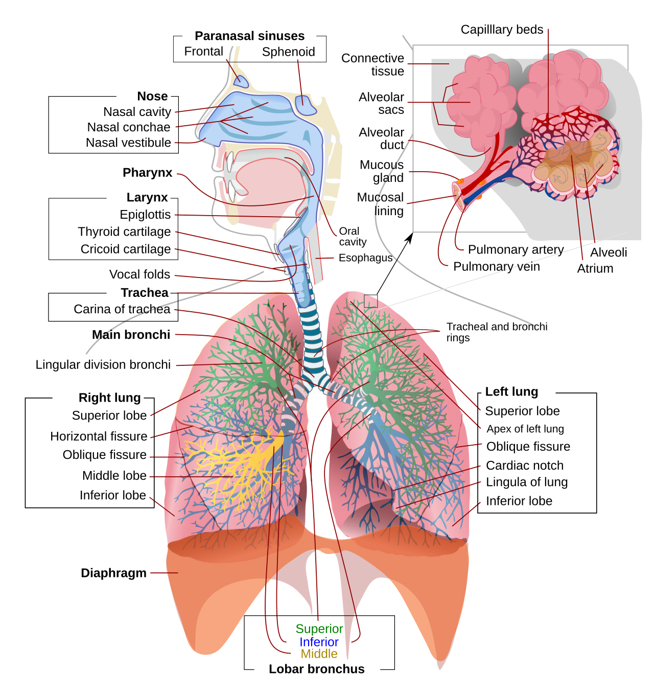
The lungs are flexible, soft, and pale pink organs located as a pair in the chest, just above the diaphragm. Interestingly, the right lung is slightly smaller but wider than the left—because the left lung is divided into two lobes while the right lung is divided into three (the left lung is smaller to allow room for the heart).
The lungs are surrounded by a double membrane—the pleura—filled with pleural fluid. This fluid reduces friction during lung movement, much like oil reduces friction between machine parts.
Pulmonary Volumes & Capacities
Various volumes and capacities have been defined to measure lung function, which are measured with a spirometer:
| Volume/Capacity | Definition | average value |
|---|---|---|
| Tidal Volume | the amount of air that moves in and out during normal breathing | ≈ 500 mL |
| Inspiratory Reserve Volume (IRV) | the amount of air that can be breathed in after normal breathing | 2000–3000 mL |
| expiratory reserve volume (ERV) | forced exhalation of air after normal exhalation | 1000–1100 mL |
| Residual Volume (RV) | air remaining in the lungs even at maximum exhalation | 1000–1500 mL |
| Vital Capacity | Maximum intake and maximum exhaust (IRV+TV+ERV) | 3400–4800 mL |
| Total lung capacity (TLC) | Maximum air the lungs can hold (VC+RV) | 5500–6000 mL |
Smoking vs. Athletes – The Truth About Lung Capacity:The vital capacity of the lungs is significantly reduced in smokers and tuberculosis (TB patients), while it is relatively high in athletes and singers – meaning the lungs, like muscles, 'adapt to use'.
◆ Pleura ◆ pleural fluid ◆ tidal volume ◆ Vital Capacity ◆ spirometer
9 Respiratory mechanisms — inhalation and exhalation
Breathing occurs in three stages—pulmonary ventilation (inhalation/exhalation), gas exchange between the alveoli and tissues, and the transport of O₂ and CO₂ through the blood. Let's understand the mechanics of breathing.
Inspiration – the active process
This is an active process, initiated by contraction of the diaphragm and external intercostal muscles. When the diaphragm contracts, it flattens and descends; simultaneously, contraction of the external intercostal muscles pulls the ribs upward and outward. Both of these actions increase the volume of the thoracic cavity—causing the air pressure in the lungs to fall below atmospheric pressure, and air rapidly fills the alveoli.
Expiration – passive process
At rest, this is a passive process. The diaphragm and external intercostal muscles relax—the ribs descend under their weight, and the diaphragm rises, forming a dome again. This reduces the volume of the chest cavity, air pressure exceeds atmospheric pressure, and air is forced out of the alveoli. In forced breathing, additional intercostal and abdominal muscles also play an active role.
The normal resting respiratory rate is about 12–16 (some sources state 16–20) times per minute, and about 600–800 mL of air is exchanged per minute. This entire mechanism is based on a pressure gradient—where higher-pressure air (the atmosphere) flows naturally toward lower-pressure areas (the lungs), much like water flows from a height downward.
| 🫁 Inspiration | 💨 Expiration |
|---|---|
| ◆ Active process ◆ Diaphragm and external intercostal muscles contracted ◆ The volume of the chest cavity increases ◆ air pressure in the lungs decreases ◆ Air enters | ◆ Passive process ◆ Muscles relax, ribs drop ◆ Decreases in the volume of the chest cavity ◆ increases air pressure in the lungs ◆ air comes out |
◆ Diaphragm ◆ external intercostal muscles ◆ Exhalation ◆ exhalation ◆ pressure gradient
10 Exchange and transport of gases
Gas Exchange
Exchange of gases occurs by diffusion due to difference in partial pressure. In the alveoli, the partial pressure of O₂ (Po₂) is approximately 105 mm Hg and that of CO₂ (Pco₂) is approximately 40 mm Hg, whereas in blood (coming from veins), Po₂ is approximately 40 mm Hg and Pco₂ is approximately 46 mm Hg. Due to this pressure difference, O₂ diffuses from the alveoli to the blood and CO₂ from the blood to the alveoli – this is called external respiration. The opposite exchange occurs in the tissues, which is called internal respiration.
Transport of oxygen
Approximately 97% of O₂ is conjugated with hemoglobin and transported as oxyhemoglobin (Hb₄(O₂)₄), leaving only 3% dissolved in plasma. The higher the Po₂, the greater the saturation of hemoglobin—the curve representing this relationship is S-shaped (sigmoid).
Hemoglobin traps O₂ where there is excess O₂ (in the lungs), and releases it where there is low O₂/high CO₂ (in working muscles) (Bohr Effect) – it is a smart mechanism that adjusts the body's supply to local demand without any central command.
Transport of carbon dioxide
CO₂ is transported in three forms:
- As bicarbonate (about 70–75%)—the largest portion; the carbonic anhydrase enzyme in RBCs converts CO₂+H₂O to carbonic acid (H₂CO₃), which dissociates into H⁺ and HCO₃⁻.
- As carbamino-hemoglobin (about 20-23%) – CO₂ binds to the NH₂ group of hemoglobin.
- In plasma in dissolved form (about 5-7%).
Chloride Shift: When HCO₃⁻ moves out of the blood cell into the plasma, Cl⁻ ions from the plasma enter the blood cell to balance it. Haldane Effect: More CO₂ is removed from the blood than O₂-Hb combination in the lungs.
| gas | Mode of transport | Percentage |
|---|---|---|
| O₂ | With hemoglobin (oxyhemoglobin) | 97% |
| O₂ | dissolved in plasma | 3% |
| CO₂ | as bicarbonate (HCO₃⁻) | 70–75% |
| CO₂ | as carbamino-hemoglobin | 20–23% |
| CO₂ | dissolved in plasma | 5–7% |
◆ partial pressure ◆ oxyhemoglobin ◆ Bohr effect ◆ bicarbonate ◆ carbonic anhydrase ◆ Chloride displacement
11 Regulation of respiration and diseases of the respiratory system
Respiration is an involuntary action controlled by respiratory centers located in the medulla oblongata and pons—the dorsal respiratory center (controls expiration), the pneumotaxic center (controls expiration and limits expiration), and the ventral respiratory center (assists in forced breathing). An increase in CO₂ and H⁺ ions in the blood increases the respiratory rate.
No matter how hard you try, you can't hold your breath for too long—because the medulla oblongata automatically 'auto-restarts' once blood CO₂ levels cross a certain threshold. This shows that even the body's most basic life processes are subject to an automatic safety mechanism, beyond voluntary control.
Major diseases of the respiratory system
| Disease | Reason | Symptoms/Prevention |
|---|---|---|
| Respiratory Asthma | Allergic reaction to external allergens | Difficulty breathing, narrowing of the bronchioles due to excess mucus; best prevention is avoidance of allergens |
| Bronchitis | Infections, smoking, air pollution | Regular cough with greenish-blue mucus; smoke and dust aversion |
| Pneumonia | bacterial infection in the alveoli | Fever, aches, severe cough; avoidance of crowds |
| Tuberculosis (TB) | Bacterial infection spread by droplets | Weight loss, cough, fever; prevented with BCG vaccine and a nutritious diet |
| Emphysema | Excessive smoking, chronic bronchitis | The elasticity of the alveoli is permanently lost; quitting smoking is the only solution. |
Smoking causes a diminished sense of smell and taste, smoker's cough, gastric ulcers, increased heart rate and blood pressure, premature wrinkles, heart disease, and stroke—and can lead to cancers of the mouth, larynx, pharynx, esophagus, lungs, pancreas, and bladder. This shows that the effects of smoking are not limited to the lungs, but extend to dozens of organs in the body.
◆ respiratory center ◆ Medulla oblongata ◆ Pneumotaxic Center ◆ Asthma ◆ emphysema ◆ BCG vaccine
12 Circulatory system – composition of blood, blood groups and coagulation
So far, we've seen how food is digested and oxygen enters the body—but who delivers these two nutrients to every corner of the body? This responsibility is performed by the circulatory system, whose central pump is the heart, and whose carrier fluid is blood.
Composition of Blood
Blood is a red, thick, slightly alkaline fluid connective tissue, which is made up of two parts – plasma (55%) and formed cells (45%).
Plasma — A pale yellow liquid composed of water (~90%), proteins (albumin, globulin, fibrinogen), and salts. Its functions include transporting digestive products and hormones, transporting waste products to the excretory organs, evenly distributing body heat, and supporting blood clotting and immunity.
| Base | RBC (erythrocyte) | WBC (leukocyte) | Platelets |
|---|---|---|---|
| Nucleus | absent | Present | absent |
| Number/mm³ | 45–55 lakhs | 4000–11000 | 1.5–4 lakhs |
| Manufacturing location | red bone marrow | bone marrow and lymphatic tissue | Bone marrow (megakaryocyte) |
| Shape | Biconcave | Irregular | small plate-like |
| Average age | ~120 days | a few days | 7–10 days |
| Main functions | Transport of O₂ and CO₂ (hemoglobin) | immunity, germicide | blood coagulation |
RBCs are formed in the red bone marrow and are destroyed in the spleen and liver after a lifespan of approximately 120 days – hence the spleen is also known as the 'graveyard of RBCs'. WBCs are of two types – granulocytes (neutrophils, eosinophils, basophils) and agranulocytes (lymphocytes, monocytes); they serve as the body's immune system.
Blood Groups – ABO System
There are four blood groups – A, B, AB and O, based on the antigens (A, B, both, or none) on the RBC membrane and the presence of antibodies in the plasma.
| Recipient Group | safe donor group |
|---|---|
| THE | Only O (universal donor) |
| A | O, A |
| B | O, B |
| AB | O, A, B, AB (universal recipients) |
O group = universal donor (no antigen); AB group = universal recipient (no antibodies). The Rh factor is also important—it determines whether the fetus is Rh+ or Rh–; incompatibility between a pregnant Rh– mother and an Rh+ fetus can lead to serious complications.
Blood clotting system
📐 Steps of Blood Coagulation – Formula/Process: Platelets at the site of injury → release thromboplastin Thromboplastin + prothrombin --Ca²⁺--> thrombin Thrombin + Fibrinogen → Fibrin (insoluble filaments) Fibrin + RBC → Blood clot
Haemophilia – A genetic disease, mainly in males, in which the blood does not clot due to a deficiency of coagulation factors and bleeding continues.
◆ Plasma ◆ RBC/WBC/Platelets ◆ Blood group (ABO) ◆ Universal donor ◆ Fibrinogen ◆ Hemophilia
13 Structure of the heart, cardiac cycle and motor system
🖼️ Internal structure of the human heart—the four chambers, valves, and major blood vessels
The heart is a muscular, hollow, red organ made of muscular tissue—about the size of a clenched fist. It is surrounded by a double-membranous sac called the pericardium, within which pericardial fluid protects the heart from external shocks and reduces friction.
four chambers and valves of the heart
The heart has two atria (small, blood-receiving chambers) at the top and two ventricles (large, powerful, pumping chambers) at the bottom. The interatrial septum and interventricular septum keep the right and left lobes completely separate—the right carries impure blood, while the left carries pure blood.
Valves allow blood to flow in only one direction—the bicuspid valve/mitral valve (left atrium-ventricle), the tricuspid valve (right atrium-ventricle), and the semilunar valves (aortic and pulmonary valves, at the ventricle-artery openings). Closing of the valves produces a "lub-dub" sound, which can be heard with a stethoscope.
Cardiac Cycle
One cardiac cycle is completed in about 0.8 seconds, consisting of diastole (relaxation) and systole (contraction). The normal adult heart rate is about 72 beats/minute, and the heart pumps about 70 mL of blood per beat (stroke volume). Thus, cardiac output (total blood pumped in one minute) = 72 × 70 ≈ 5000 mL, or about 5 liters per minute.
Conducting System of the Heart
The sequence of heartbeats is determined by a special electrical conduction system:
- Sino-atrial node (SA Node) – Located in the upper wall of the right atrium; called the natural pacemaker, it is where the heartbeat begins.
- Atrioventricular node (AV Node) — Located between the right atrium and ventricle; transmits impulses from the SA node to the ventricles.
- Bundle of His and Purkinje Fibres – They rapidly transmit impulses to the ventricular muscles and produce coordinated contractions.
If the SA node becomes faulty, an artificial pacemaker is implanted in the chest. An ECG (Electrocardiogram) is a device that records the conduction of the heartbeat, which is helpful in detecting heart-related disorders.
◆ atrium and ventricle ◆ bicuspid valve ◆ tricuspid valve ◆ Lubb-dub ◆ SA node (pacemaker) ◆ cardiac output
14 Blood Vessels and Types of Circulation
Blood Vessels
| Base | Arteries | Veins | Capillaries |
|---|---|---|---|
| Direction | away from the heart | towards the heart | connects arteries and veins |
| wall | thick, muscular, flexible | thin, less muscular | single-celled layer |
| blood type | O₂-rich (exception: pulmonary artery) | CO₂ containing (exception: pulmonary vein) | Mixed due to exchange |
| Closet | Absent (except near the heart) | present (prevents backflow) | absent |
| blood flow | fast and pulsating | slow and steady | Extremely slow (for exchange) |
Pulmonary artery and vein—exception to the name:The pulmonary artery is the only artery that carries impure blood—yet it is called an "artery" because it carries blood away from the heart. Similarly, the pulmonary vein is the only vein that carries pure blood, yet it is called a "vein" because it brings blood to the heart—that is, the names artery and vein are determined not by the purity of the blood, but by the direction of flow (from/to the heart).
Types of circulation
- Systemic Circulation—The aorta carries purified blood from the left ventricle to the rest of the body; the great veins return impure blood to the right atrium.
- Pulmonary Circulation—Impure blood from the right ventricle goes to the lungs; the pulmonary veins bring pure blood to the left atrium.
- Coronary Circulation – The blood supply to the heart's own muscles is through the coronary arteries.
- Hepatic Portal System – Nutrient-rich blood from the small intestine travels directly to the liver through the hepatic portal vein, allowing the liver to absorb and process nutrients.
In humans, blood passes through the heart twice, hence the name Double Circulation—first through the body-heart (somatic) and then the heart-lung-heart (pulmonary) cycle. This dual system ensures that pure and impure blood never mix, keeping oxygen distribution highly efficient—much like a city has separate pipelines for drinking water and wastewater.
◆ Artery vs. Vein ◆ pulmonary artery ◆ physiological circuit ◆ coronary circulation ◆ hepatic vascular system ◆ double circulation
15 Diseases of blood pressure, lymphatic system and circulatory system
Blood Pressure
The pressure that blood exerts on the walls of arteries during ventricular contraction (systole) is called blood pressure—systolic pressure (higher) and diastolic pressure (lower). The normal reading for a healthy adult is 120/80 mm Hg, measured with a sphygmomanometer. When systolic pressure ≥140 and diastolic pressure ≥90 mm Hg remain persistent, it is called hypertension—it can adversely affect the heart, brain, and kidneys.
Lymphatic System
The lymphatic system is a complementary system to the circulatory system. It consists of lymph (a colorless fluid containing plasma and WBCs but devoid of RBCs), lymph vessels, lymph nodes, and lymphoid organs (spleen, thymus, bone marrow). The main functions of the lymphatic system are to collect excess fluid from tissues and return it to the blood, aid in the absorption of fats (via lacteals), provide immunity, and prevent the spread of infection.
Lymph flows in only one direction—from the organs to the heart, never the other way around—and has no pumping mechanism; it is propelled forward by muscle movement.
Major diseases of the circulatory system
- Atherosclerosis – Plaque buildup due to cholesterol deposits on the artery walls, gradually narrowing and hardening the arteries.
- Heart attack — a clot (thrombosis) in a coronary artery that blocks blood supply to the heart muscle; obesity, smoking, high blood pressure, and high cholesterol are major risk factors.
- Stroke – Blockage or clot in the arteries of the brain.
- Anemia – a deficiency of hemoglobin; caused by a deficiency of iron and vitamin B12.
- Varicose Veins – The veins in the legs become stretched and crooked in people who stand for long periods of time.
- Leukemia (blood cancer) — uncontrolled production of WBCs.
Arteriosclerosis—connections to the previous chapter:Remember—in the previous chapter, we learned that a diet high in fat and cholesterol can cause plaque to build up in the arteries. This process, in turn, can lead to arterial hardening and heart attacks—meaning that diet and nutrition are directly linked to the health of the circulatory system.
◆ Blood pressure (120/80) ◆ sphygmomanometer ◆ lymphatic system ◆ arterial stiffness ◆ Heart attack ◆ Anemia
16 Excretory System – Nitrogenous Wastes and the Structure of the Kidneys
The digestive, respiratory, and circulatory systems together provide the body with nutrition and energy—but who cleans up the waste generated during this process? This responsibility lies with the excretory system, which maintains homeostasis by removing waste products produced by the body's cells.
Three types of nitrogenous waste
| Type | Toxicity | Example organisms |
|---|---|---|
| Ammonia (Ammoniolytic) | Most toxic, requires a lot of water | Most fishes, aquatic insects, amoebas |
| Urea (Ureotelic) | Moderately toxic; liver converts ammonia to urea | humans and other mammals |
| Uric acid (Uricotelic) | Least toxic, in paste/pellet form | Birds, reptiles, insects |
💡 How do birds stay poison-free inside their eggs?: Birds are uricotelic, so embryos can accumulate nitrogenous wastes in the form of uric acid (a less toxic solid) while developing in the enclosed environment of the egg without harming themselves—whereas accumulation of the more toxic ammonia would kill the embryo. This is a brilliant adaptation strategy by nature.
parts of the human excretory system
Two kidneys (Kidneys — main organs, filter blood and form urine), two ureters (Ureters — carry urine to the bladder), a urinary bladder (Temporary storage of urine) and a urethra (Urethra — the way for urine to exit the body).
🖼️ Structure of the nephron – the functional unit of urine formation
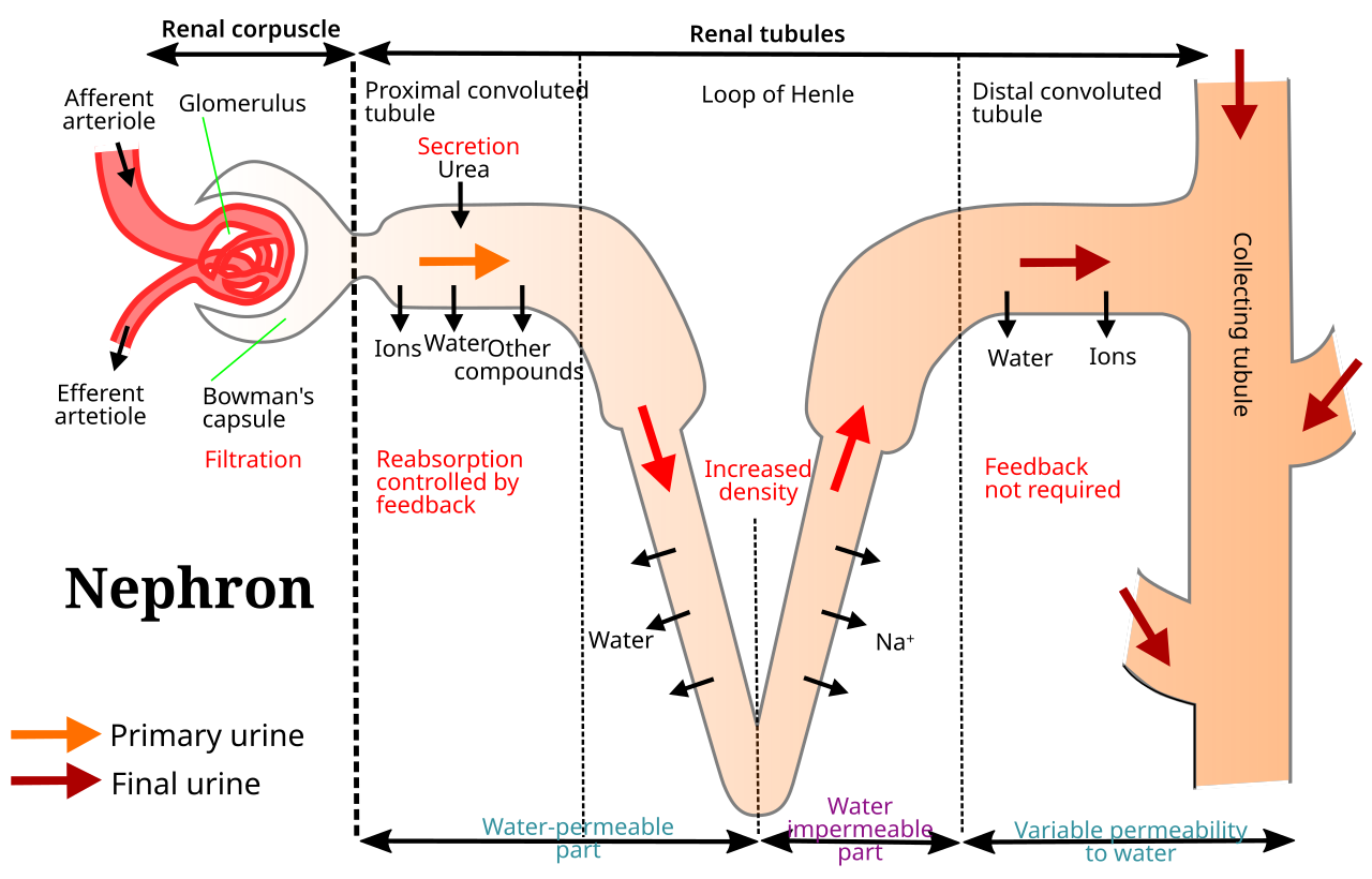
structure of the kidney
The kidney is a bean-sized, red organ—10–12 cm long, 6 cm wide, and 3–4 cm thick. It is located in the abdominal cavity, beneath the diaphragm, on either side of the vertebral column. The kidney has a groove—the hilum—on its medial surface, through which the ureters, nerves, and blood vessels enter.
In a vertical section of the kidney, two parts are clearly visible – the cortex (Cortex, the outer part, appears granular due to Malpighian corpuscles) and the medulla (Medulla, the inner part, divided into triangular pyramids, between which the renal columns of Bertini are embedded).
◆ amniotic ◆ ureterocele ◆ Uricosurgeon ◆ Hylam ◆ Renal Cortex ◆ Renal medulla
17 Nephron and the three-step process of urine formation
Each kidney contains 10-12 lakh fine, long and coiled tubes called nephrons – this is the structural and functional unit of the kidney.
Parts of the nephron (in order)
- Malpighian Corpuscle — Bowman's Capsule + Glomerulus. The afferent arteriole brings blood, the efferent arteriole takes it back. The inner wall of Bowman's capsule is made up of podocytes, which act as a highly permeable sieve.
- Proximal convoluted tubule (PCT)—Long, thick tubule; most reabsorption occurs here.
- Loop of Henle—U-shaped portion located in the medulla; composed of a descending and ascending arm.
- Distal convoluted tubule (DCT) — Located in the cortex, opens into the collecting duct.
- Collecting Duct—Located in the pyramids of the medulla; several collecting ducts join to form the duct of Bellini, which ultimately opens into the renal pelvis.
Each kidney filters about 180 liters of fluid daily—that is, both kidneys together filter the equivalent of a small swimming pool—but the final urine produced is only 1-2 liters, because the body reabsorbs more than 99% of the water. This shows how efficient the kidneys are as recycling plants.
three stages of urine formation
- Ultrafiltration – The glomerulus has numerous tiny pores in its walls; the hydrostatic pressure of the blood (~70 mm Hg) filters water, glucose, amino acids, and urea into Bowman's capsule (blood cells and larger proteins are not filtered). This fluid is called the glomerular filtrate.
- Selective Reabsorption—Approximately 65–80% of the PCT (glucose, amino acids, salts) is returned to the blood by active transport; water-salt balance is maintained by a countercurrent mechanism in the loop of Henle; final water reabsorption is achieved in the DCT.
- Tubular Secretion — The cells of the PCT and DCT release K+, H+, ammonia, uric acid, and some drugs into the tubules through active transport to maintain blood homeostasis. The filtrate is then called urine.
The maximum concentration of a substance in the blood at which it is completely reabsorbed from the glomerular filtrate. High-threshold substances such as glucose are normally completely reabsorbed—that is why glucose is not found in the urine of a healthy person; in diabetes, blood sugar rises so high that the threshold value is exceeded and glucose appears in the urine.
Composition of urine: Color light yellow (due to urochrome pigment), quantity 1-2 liters per day, pH slightly acidic (about 6), water 96%, urea about 2%, and other organic and inorganic salts.
◆ What is the name of the place? ◆ glomerulus ◆ Podocytes ◆ ultrafiltration ◆ selective reabsorption ◆ Tubular secretion
18 Hormonal regulation, dialysis and diseases of the excretory system
Hormonal regulation (osmoregulation)
Kidney function is primarily controlled by two hormones:
- Aldosterone—secreted from the adrenal cortex; increases Na+ reabsorption in the renal tubules, maintaining sodium and water balance in the body.
- Vasopressin/ADH (Anti-Diuretic Hormone) — secreted by the posterior pituitary gland; determines urine concentration by controlling water reabsorption in the collecting duct — when water is deficient, more ADH is secreted, resulting in less and more concentrated urine.
Dialysis
When the kidneys do not function properly, urea in the blood increases (uremia). The process of removing urea from the blood using an artificial kidney is called hemodialysis. The patient's blood is passed through tubes made of cellophane membrane, which allows small molecules (urea, uric acid) to pass through but blocks larger protein molecules. The concentration gradient forces harmful substances from the blood into the dialyzing fluid, and the purified blood is then returned to the patient's vein.
💡 Cellophane membrane — body-mimicking technology: The cellophane membrane of a dialysis machine works just like the glomerulus/podocytes of a real kidney—letting small molecules pass through but blocking larger protein molecules. This shows that modern medical technology is often created by mimicking nature's own 'filter design.'
Major diseases of the excretory system
| Disease | Causes/Symptoms |
|---|---|
| Uraemia | Excessive urea in the blood; fatal in severe cases |
| Gout | Excessive uric acid in the blood causing accumulation in the joints |
| Kidney Stones | Calcium oxalate, phosphate, and uric acid crystal deposits |
| Nephritis | Inflammation of the glomerulus due to bacterial infection, blood/protein in the urine |
| glycosuria | Glucose in the urine – a sign of diabetes mellitus |
| Diabetes insipidus | Excessively dilute urine due to ADH deficiency |
Because one kidney can meet the body's needs (which is why living donors can donate a kidney), kidney disease often remains asymptomatic until 70–80% of the kidney function is lost—that's why regular health checkups and adequate hydration are essential for kidney health.
◆ Aldosterone ◆ ADH/Vasopressin ◆ hemodialysis ◆ Uremia ◆ Gout ◆ nephritis
• The digestive system includes the alimentary canal (8–10 m long, from the mouth to the anus) and three digestive glands (salivary glands, liver, and pancreas); carbohydrates, proteins, and fats are broken down stepwise into simpler absorbable units by amylase, pepsin–trypsin, and lipase–bile salts, respectively. • The villi and microvilli of the small intestine increase the absorptive surface by 1000 times; glucose and amino acids are absorbed into the blood capillaries and fatty acids into the lymphatic lacteals; hormones such as gastrin, secretin, and CCK regulate the secretion of digestive juices. • The respiratory system delivers air from the nose to the alveoli; Expiratory (mesenchymal contraction, active) and expiratory (relaxation, passive) pressures work on the principle of gradient; O2 is transported 97% from hemoglobin and CO2 mainly (70-75%) in bicarbonate form. • Respiration is regulated involuntarily by the respiratory centers in the medulla oblongata based on blood CO₂ levels; clean air and vaccination are essential to prevent diseases such as asthma, bronchitis, pneumonia, and tuberculosis. • The circulatory system consists of the blood (plasma + RBCs/WBCs/platelets), the heart (four chambers, valves, SA node pacemaker), and blood vessels (arteries, veins, and capillaries); the cardiac cycle is ~0.8 seconds, and cardiac output is ~5 liters/minute. • In humans, double circulation (systemic + pulmonary) passes blood through the heart twice; blood pressure is normally 120/80 mm Hg; the lymphatic system helps absorb excess tissue fluid and fat; arterial strictures and heart attacks are major cholesterol-related diseases. • The excretory system removes nitrogenous wastes (mainly urea in humans) through the kidneys; nephrons (10-12 lakh per kidney) convert 180 litres of fluid into only 1-2 litres of urine daily through a three-step process of ultrafiltration, selective reabsorption and tubular secretion. • The hormones ADH and aldosterone regulate water-salt balance; kidney failure requires dialysis or transplantation; the four systems—digestion, respiration, circulation, and excretion—work together to run the body's harmonious, automatic life cycle.