- Introduction to the cell, cell theory and history of discovery
- Shape and size of the cell and unicellular and multicellular organisms
- prokaryotic and eukaryotic cells
- Cell Wall
- Cell Membrane and Fluid Mosaic Model
- Mitochondria—the powerhouse of the cell
- Plastids
- endoplasmic reticulum and golgi bodies
- Ribosomes, centrosomes and microtubules
- Cilia, flagella, vacuole, microbodies, lysosomes and spherosomes
- Nucleus
- Chromosomes
- Cell Cycle and Mitosis
- Meiosis
- Difference between plant and animal cells
- Major discoveries in cytology
On a quiet afternoon in 1665, in the laboratory of the Royal Society of London, an inquisitive scientist named Robert Hooke sat in front of a hand-made compound microscope. He cut a very thin slice of cork (oak bark), used for making bottle caps, and placed it under a glass. As he peered into the microscope, his eyes widened—he saw thousands of tiny, tightly packed, empty compartment-like structures on the surface of the cork, much like the tiny chambers in a beehive or the many similar rooms in a large hostel. Hooke named these empty compartments the Latin word "cellula," meaning "little room," which later became "cell" in English. He wrote a detailed description, complete with illustrations, in his book "Micrographia." Interestingly, what Hooke saw was actually just the wall of dead cells, or cell walls, not living cells—yet his discovery laid the foundation for one of the greatest revolutions in biology. Today we know that our entire body—bone, skin, muscle, brain, everything—is made up of millions and billions of these tiny "compartments," which we call cells. Let's understand the entire world of this smallest room in detail.
1 Introduction to the cell, cell theory and history of discovery
Just as a large building is made up of small bricks, every living thing—be it a tiny bacterium or a giant elephant—is made up of tiny "biological bricks." We call these biological bricks cells. A cell is the smallest, fertile (capable of forming new cells of its own), and membrane-enclosed structural and functional unit of a living organism. In other words, imagining life without cells is like imagining a house without bricks.
The protoplasm found within the cell is called the "physical basis of life," because it is in it that all the chemical and physical reactions of life take place. DNA (deoxyribonucleic acid), the carrier of genetic traits, is located within the cell, which is also called the "master molecule" of life because it is this molecule that determines how an organism will look and function. The study of the structure, organization, and functioning of the cell is called cytology.
The interesting history of the discovery of the cell
The discovery of the cell wasn't a one-day event, but rather the result of a long scientific journey spanning nearly two hundred years. Let's explore this journey step by step.
- Robert Hooke (1665) — As mentioned in our story above, Hooke first observed cell-like structures in slices of cork and named them 'cellula.' He actually observed dead cell walls, not living cells.
- Antonie van Leeuwenhoek (1674 AD) — Dutch scientist Leeuwenhoek created a highly powerful single-lens microscope with his own hands and is credited with being the first person in history to observe a living cell. He observed and drew bacteria, red blood cells, sperm, and unicellular organisms (such as protozoa) floating in pond water. Leeuwenhoek is therefore also known as the "Father of Microbiology."
- Carl (Corti, 1772) — was the first scientist to observe protoplasm.
- Robert Brown (1831) — Scottish botanist Robert Brown discovered the nucleus while studying orchid root cells. This was a pivotal discovery in cell biology because the nucleus controls all of the cell's activities.
- Purkinje (1840) – He named the fluid inside the cell as 'protoplasm'.
- Matthias Schleiden (1838) – German botanist Schleiden, after studying the cells of various plants under a microscope, concluded that the body of all plants is made up of cells.
- Theodor Schwann (1839) — German zoologist Schwann expanded on Schleiden's findings, demonstrating that animal bodies are also composed of cells. Together, Schleiden and Schwann formulated the Cell Theory in 1838-39.
- Rudolf Virchow (1855) — The German scientist Virchow added a very important modification to the cell theory. He made the famous statement in Latin—'Omnis Cellula e Cellula,' meaning 'Every cell is formed from a previous cell.' This simply means that new cells do not arise spontaneously from thin air, but always arise from the division of a living, pre-existing cell.
Remember – the father of modern cell scienceC. P. Schwanson(C.P. Swanson) is called, whileA. K. Sharma(A.K. Sharma) is considered the father of Indian cytology – this fact is useful to remember from the RAS exam point of view.
Modern Cell Theory
Over time, scientists added Virchow's contributions to Schleiden-Schwann's original theory, creating the modern cell theory, which is still considered the foundation of biology today:
- The body of all living things is made up of cells and their products.
- New cells are always formed by the division of preexisting cells, not spontaneously.
- The cell is the structural and functional unit of the living body – that is, the body is also made up of cells and all the functions of the body are also performed at the cell level.
- Each cell contains the organism's genetic material (in the form of DNA), which is passed on to daughter cells.
- Each cell represents the continuity of life—that is, it continues to grow from generation to generation, maintaining its origin and basic nature.
💡 Everyday Example - Understand Cell Theory in Simple Language: Just as new flats in a large society are built not by demolishing existing ones but by building new ones based on the same design—so too, a new cell does not suddenly arise out of thin air, but always through the division of an old, living cell. This is the essence of Virchow's statement, "Omnis Cellula e Cellula."
Viruses – an exception to the cell theory
The structure of viruses cannot be explained by the cell theory, so they are considered exceptions to the cell theory. The main reasons for this are the following:
- Viruses do not have the ability to divide independently – they require a living host cell for their multiplication.
- Plasma membrane, cytoplasm, enzymes and other cellular components are absent in them.
- Only one type of nucleic acid – either DNA or RNA – is found in them, whereas in living cells both types of nucleic acids are present together.
That is why viruses are often called the 'link between living and non-living' – outside the host cell they behave like lifeless crystals, but as soon as they enter the host cell they start multiplying like living organisms.
| Scientist (Year) | Major contributions |
|---|---|
| Robert Hooke (1665) | Observed cell wall in cork, named it 'Cell' |
| Leeuwenhoek (1674) | First observed living cell |
| Robert Brown (1831) | Discovery of the Nucleus |
| Purkinje (1840) | named protoplasm |
| Schleiden (1838) | The body of all plants is made up of cells - propounded |
| Dog (1839) | The body of all animals is made up of cells - proposed |
| Rudolf Virchow (1855) | 'Omnis Cellula e Cellula' - Modification of the Cell Theory |
◆ Cell ◆ Cytology ◆ Protoplasm ◆ Cell Theory ◆ Nucleus ◆ Virus ◆ Cellula
2 Cell size and shape and unicellular and multicellular organisms
Have you ever wondered how small the world's smallest cell can be, and how big the largest cell? The size and shape of cells depend on the function they perform—just as different shaped tools are made in a factory for different jobs. In unicellular organisms, cells can be rod-shaped, spiral, comma-like, or a variety of other shapes. Plant cells typically range from 15 to 100 micrometers (μm) in diameter.
The smallest cell is considered to be Mycoplasma or PPLO (Pleuro Pneumonia Like Organism) – which has a diameter of only about 0.1 micrometer, meaning it is so small that it is barely visible even with a simple microscope. In contrast, the largest cell is an ostrich egg cell, which has a diameter of about 15 centimeters – meaning you can see an entire cell in the palm of your hand with just one eye! The longest unicellular plant cell is that of an alga called Acetabularia, which is about 10 centimeters long, while the longest fibers among multicellular plants are those of Boehmeria nivea (Boehmeria nivea) are found in plants, and can range from 22 to 55 centimeters in length. Neurons in the human body can also be up to a meter long—such as the nerve cells running from the spinal cord to the big toe.
unicellular and multicellular organisms
On the basis of complexity of structure, living organisms are divided into two classes - Unicellular Organisms, whose entire body is made up of only one cell, like Amoeba, Paramecium and bacteria; and Multicellular Organisms, whose body is made up of many cells, like humans, animals, birds and plants. In unicellular organisms, that one cell alone performs all the functions like food intake, respiration, excretion and reproduction, whereas in multicellular organisms, different cells are divided for different specific functions - this is called 'Division of Labour'.
| Base | unicellular organisms | multicellular organisms |
|---|---|---|
| Number of cells | only one cell | many cells |
| Structure | Simple | Complex |
| division of work | No, one cell does all the work | There is a clear division of work |
| size | Usually microscopic | Usually large (macroscopic) |
| tissues and organs | absent | present (tissue, organ, organ system) |
| Dependency | A cell lives independently | cells depend on each other |
| Growth | increase in cell size | increase in the number of cells |
| Example | Amoeba, Paramecium, Bacteria | humans, animals, plants |
| 🔵 Examples of unicellular organisms | 🔴 Examples of multicellular organisms |
|---|---|
| ◆ Amoeba ◆ Paramecium ◆ Chlorella – unicellular algae ◆ Bacteria ◆ Euglena | ◆ Humans and all mammals ◆ trees and flowering plants ◆ Reptiles and birds ◆ aquatic animals like fish ◆ Fungi (multicellular type) |
◆ unicellular organisms ◆ multicellular organisms ◆ micrometer (μm) ◆ Mycoplasma (PPLO) ◆ division of work
3 Prokaryotic and eukaryotic cells
Based on the development and organization of the nucleus, all the cells of the world have been divided into two major classes—prokaryotic cells and eukaryotic cells. This classification is considered the most fundamental classification of biology, because it determines how simple or complex the internal structure of an organism is.
Prokaryotic Cell
The word 'Prokaryotic' is made up of two Greek words –Pro(which means primitive) andKaryon(Which means nucleus). These types of cells lack a membrane-bound well-organized nucleus (True Nucleus).
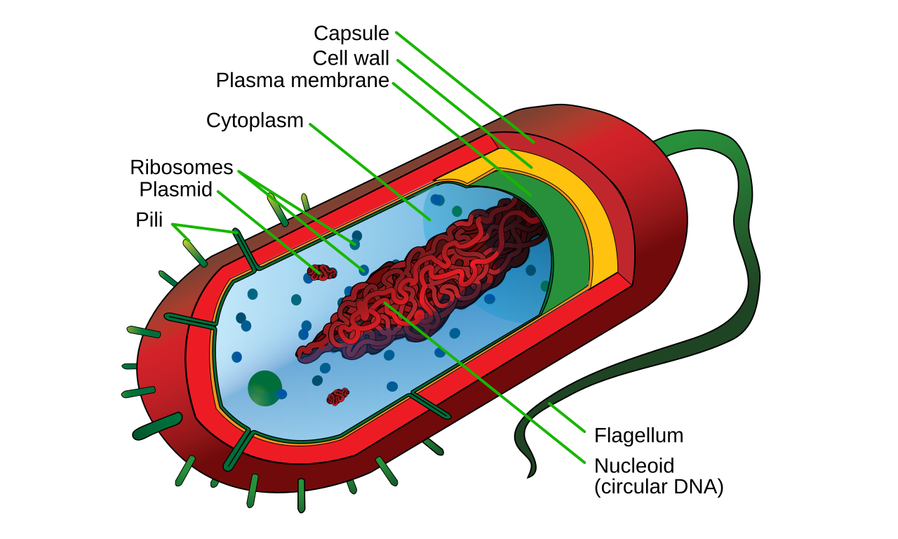
The cells of organisms such as Blue-Green Algae (Cyanobacteria), Bacteria, and Mycoplasma completely lack a well-organized membrane-bound nucleus and double-layered membrane-bound organelles (such as mitochondria, Golgi bodies). The nucleus-equivalent region in such cells is called a 'prokaryon' or 'nucleoid', where the genetic material lies freely, not in the form of chromosomes, but as a naked, circular, coiled nucleic acid (DNA). They also lack histone proteins and do not undergo mitosis—instead, they reproduce by binary fission.
Example: Members of the kingdom Monera such as True Bacteria, Cyanobacteria (Blue-wave Algae), Archaebacteria, Mycoplasma and Actinomycetes.Escherichia coli(Normal bacteria found in our large intestine).
Eukaryotic Cell
'Eu' means well and 'karyon' means nucleus. These cells have a well-organized, membrane-bound, distinct nucleus, which is why they are also called karyocytic cells.
Eukaryotic cells contain several organelles with double membranes, such as mitochondria, chloroplasts, Golgi bodies, and the endoplasmic reticulum. The nucleus contains one or more nucleoli. Their genetic material is organized in the form of chromosomes, which are composed of histone and non-histone proteins. They undergo both mitosis and meiosis. Most animal and plant cells fall into this category, such as humans, amoebas (except for some exceptions, protists, where amoebas are also eukaryotic), fungi, and all plant cells.
🖼️ Internal structure of a typical prokaryotic cell (bacterial cell)
| Basis of comparison | prokaryotic cell | eukaryotic cell |
|---|---|---|
| Size and growth | Small and Primitive | large and developed |
| well-organized nucleus | Absent (nucleoid only) | present, surrounded by a nuclear membrane |
| genetic material | Naked, circular DNA (without histones) | as chromosomes (including histones) |
| respiratory site | mesosome (folding of the cell membrane) | mitochondria |
| Membrane-bound organelles (ER, Golgi apparatus, lysosomes) | absent | Present |
| ribosomes | Type 70S only | Both 70S (in mitochondria/chloroplasts) and 80S |
| microtubules and microfilaments | absent | Present |
| centrosome | absent | Present in most animal and some plant cells |
| cell division | Simple Binary Fission | Mitosis and Meiosis |
| cell wall composition | Made of mucopeptide/peptidoglycan | Cellulose present in plants, absent in animals |
| Example | Bacteria, blue-green algae, mycoplasma | Plants, Animals, Fungi, Protists |
A prokaryotic cell can be compared to a one-room house, with a kitchen, bedroom, and study—all in one open room, without any walls or partitions. In contrast, a eukaryotic cell is like a large, well-organized mansion, with separate rooms (organelles) enclosed by separate walls for each function—such as the kitchen (mitochondria, where energy is 'cooked'), the storeroom (vacuole), and the main control room (nucleus).
◆ prokaryotic cell ◆ eukaryotic cell ◆ nucleoid ◆ Mesosomes ◆ 70S ribosome ◆ 80S ribosome ◆ Bifurcation
4 Cell Wall
Plant cells require a rigid outer covering—called the cell wall—to maintain their definite shape and provide protection from external shocks. This is a strong, non-living layer found just outside the cell membrane. Note: Animal cells do not have cell walls; only plants, fungi, and bacteria possess this structure, and its chemical composition varies among these three.
For example, the cell wall of fungi is made of the polysaccharide chitin, or fungal cellulose, while that of algae is made of cellulose and pectin. In bacteria, it is made of peptidoglycan (mucopeptide)—this is why antibiotics like penicillin destroy this cell wall, killing them.
Organization of the cell wall (four layers)
A. Middle Lamella – Made of calcium pectate, this is the first, outermost layer that connects two adjacent cells.
B. Primary Wall – This is a thin and flexible layer, which keeps expanding as the cell grows.
C. Secondary Wall – This thick and tough layer is formed when the growth of the cell stops, it gives extra strength to the cell.
D. Tertiary Wall – The innermost, extra strong layer found in some specialized cells (such as wood cells).
In the final stage of cell division (telophase), the Golgi apparatus, endoplasmic reticulum, and vesicles combine to form a structure called the cell plate or phragmoplast – this further forms the middle lamella, on which the primary, secondary, and tertiary wall layers are deposited, forming the entire cell wall.
Growth and chemical changes in the cell wall
- Intussusception – When new substances accumulate between the fibres of the primary wall.
- Apposition – When new substances are deposited inward in the form of layers.
- Cellulose and pectin get deposited on the walls of the cells of the oblong tissue and make them strong.
- Lignin accumulates in the vessels and sclerenchyma cells, making the wall woody and extremely hard – this is why wood is so strong.
- The deposition of cutin on the epidermis forms a waterproof layer called the cuticle, which reduces transpiration (unnecessary loss of water) from the plant—just as this thick layer on cacti helps them conserve water in the desert.
- The deposition of suberin on cork cells makes them completely waterproof – this suberin-rich cork is used to make bottle caps, as we saw in Hooke's story.
Major functions of the cell wall
- To give a definite shape and size to the cell.
- To provide mechanical strength and toughness to the cell.
- To protect the cytoplasm and cells from adverse conditions (such as excessive water absorption).
- Transport (exchange) of intercellular substances through tiny pores called plasmodesmata.
- Maintaining the balance of osmotic pressure between cells.
When you look at onion peel under a microscope (a popular school laboratory experiment), the distinct brick-like boundaries you see are actually cell walls made of cellulose, about 1 micrometer thick—just like the cork cell walls observed by Hooke.
◆ Cell Wall ◆ middle lamella ◆ cellulose ◆ lignin ◆ cutin ◆ Suberin ◆ Phragmoplast
5 Cell Membrane and Fluid Mosaic Model
If we think of the cell wall as the outer boundary wall of a house, then the cell membrane is like the main doors and windows of that house—determining what comes in and what goes out. All cells (whether plant or animal) have a selectively permeable, electrically charged, and selective membrane surrounding the cytoplasm, composed primarily of lipids and proteins. This is also called the plasma membrane, plasma membrane, or plasmalemma.
Three historical models for understanding the structure of the cell membrane
- Danielli-Dawson's 'sandwich model' (1938) — These scientists first proposed that the membrane structure resembles a sandwich, with a layer of fat (lipid) sandwiched between two dense protein layers—like butter sandwiched between two slices of bread.
- Robertson's 'Unit Membrane Hypothesis' (1958) – Robertson stated that the membrane is a unit layer about 70-80 Å (Angstrom) thick, which is found in one or two layers in every organelle – such as double layer in chloroplast, mitochondria and nucleus, while single layer in lysosomes and vacuoles.
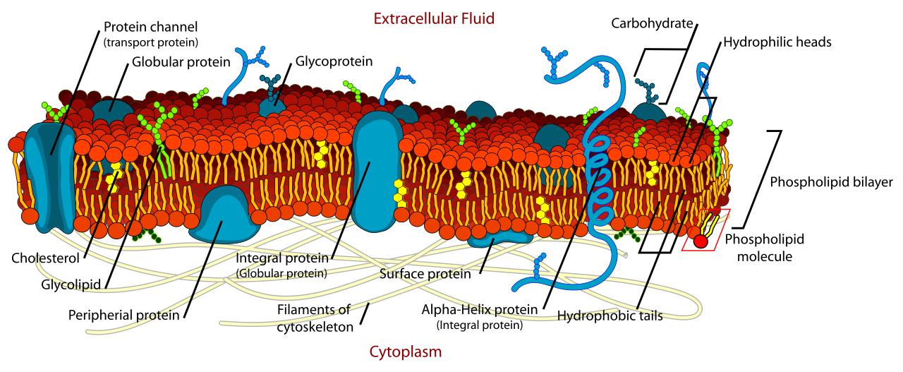
- Singer and Nicholson's "Fluid Mosaic Model" (1972)—This remains the most widely accepted model today. According to this, the cell membrane is composed of a bilayer of phospholipids, within which protein molecules "float" like pieces of a mosaic—hence the name "fluid" model.
📐 Thickness of a Liquid Mosaic Model — Formula/Procedure: Total membrane thickness ≈ 75 Å Thickness of phospholipid bilayer ≈ 45 Å 1 Å (angstrom) = 10⁻¹⁰ meters
A phospholipid molecule has two parts—a hydrophilic head, which is attracted to water, and a hydrophobic tail, which repels water. This is why the phospholipid bilayer has all its heads facing outward (in contact with water) and all its tails facing inward (facing each other). Two types of proteins adhere to it—extrinsic or peripheral proteins, which adhere to the membrane surface, and intrinsic or integral proteins, which penetrate the fatty layer and regulate the movement of substances by forming porous channels. Apart from this, cholesterol molecules are also scattered in the membrane, which control the fluidity of the membrane, and glycoprotein and glycolipid molecules present on the surface of the cell help the cell in establishing its identity (Cell Recognition) – like our blood group (Blood Group A, B, AB, O) is also determined by these surface molecules.
🖼️ Fluid mosaic model — arrangement of phospholipid bilayer and protein molecules
transport of substances across the cell membrane
The cell membrane acts as a selective membrane—it doesn't allow every substance in or out, but only selects certain ones. This movement of substances occurs primarily through two methods:
| 🟢 Passive Transport | Active Transport |
|---|---|
| ◆ Substances move from higher concentration to lower concentration ◆ No additional external energy (ATP) is required. ◆ Example – Diffusion and Osmosis ◆ For example, sugar dissolving in tea and spreading | ◆ substances move from their lower concentration to higher concentration ◆ The extra energy is expended in the form of ATP. ◆ occurs with the help of a carrier protein (permease) ◆ E.g., sodium-potassium pump in nerve cells |
Kidney dialysis machines work on this principle of osmosis—when the kidneys fail, the dialysis machine uses an artificial semi-permeable membrane to filter waste products from the blood, just as cell membranes do naturally.
Endocytosis and Exocytosis
- Endocytosis — The process of engulfing foreign substances by the cell membrane. If the substance is solid, it is called phagocytosis (such as the engulfment of bacteria by a white blood cell), and if it is liquid, it is called pinocytosis.
- Exocytosis – The process of a cell expelling unnecessary or secretory substances through its membrane – such as the release of insulin from pancreatic cells into the blood.
Special modifications of animal cell membranes
- Microvilli – The membrane in the epithelial cells of the intestine forms finger-like projections, which increase the area of absorption of nutrients many times.
- Desmosomes – Thread-like structures formed on the membrane to increase the mechanical strength of cells, whose filaments are called tonofibrils.
- Interdigitation – Extensions formed on the membrane to establish close contact between adjacent cells.
◆ cell membrane ◆ Plasmalemma ◆ Liquid mosaic model ◆ phospholipid bilayer ◆ diffusion ◆ osmosis ◆ Active transport ◆ Endocytosis ◆ Exocytosis
6 Mitochondria — the powerhouse of the cell
The word 'Mitochondria' comes from the Greek languageMyth(thread) andChondrionIt is composed of (particles). It was first discovered by Kölliker (1880), Flemming (1882) named it 'phyla' and Altmann (1894) called it 'bioplast'. Finally, C. Benda (1897) gave it the name 'mitochondria', which remains in use today.
structure of mitochondria
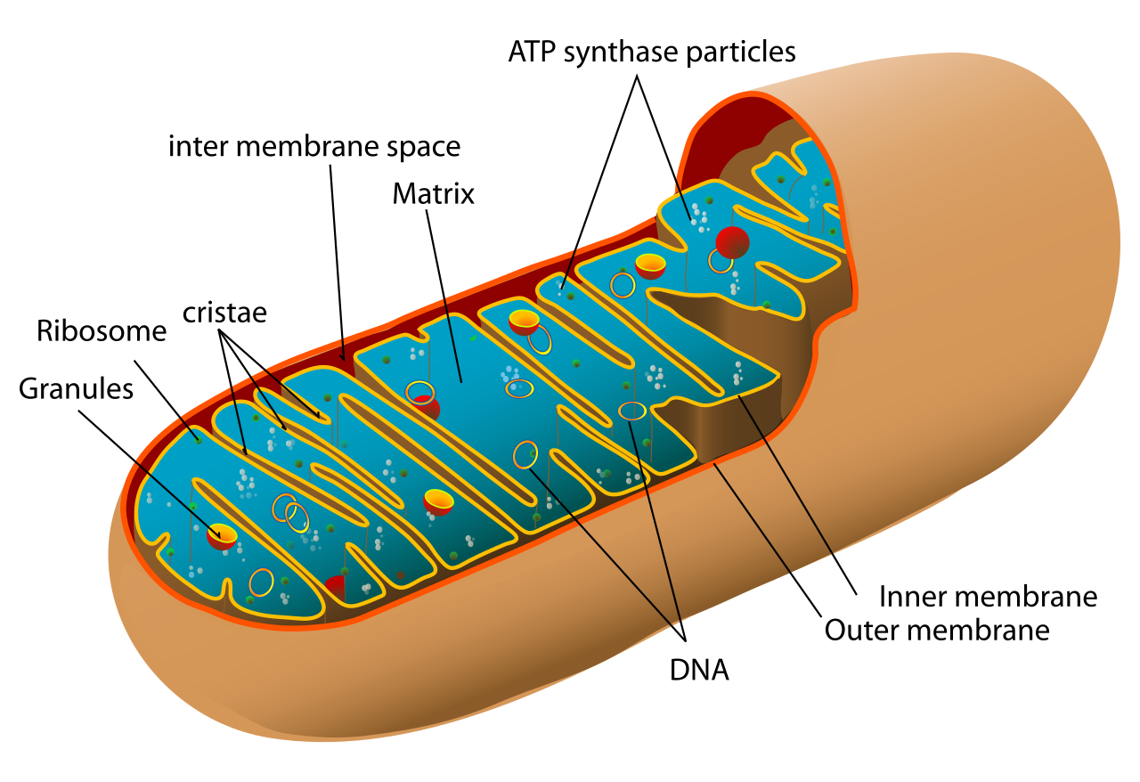
Mitochondria are organelles surrounded by a double membrane. Their outer membrane is flat, while the inner membrane bears numerous finger-like projections called cristae—these projections multiply the surface area of the inner membrane to maximize energy production. The space between the two membranes is called the perimitochondrial space, which contains various respiratory enzymes, while the innermost layer is filled with a jelly-like, protein-rich, homogeneous substance called the matrix. The surface of the cristae contains tiny particles called elementary particles, oxosomes, or F₁ particles—these are where the ATP synthetase enzyme, which performs oxidation and phosphorylation, is found.
Functions of mitochondria
Mitochondria are called the cell's "powerhouse" or "energy house" because they are where aerobic respiration occurs, storing energy from the oxidation of food (glucose) in the form of adenosine triphosphate (ATP). When the cell needs energy for any biological function, ATP is broken down into ADP, a phosphate molecule, and energy—one ATP molecule releases approximately 7.3 kilocalories of energy. After glycolysis, the glucose molecule breaks down into two pyruvic acid molecules, which are oxidized in a cyclic pathway by various respiratory enzymes present in the matrix—this is called the Krebs Cycle. The final step of this entire process is called oxidative phosphorylation, which occurs in oxosome particles located on the cristae.
Mitochondria have their own circular, naked DNA, which gives them the ability to self-replicate—but this ability is achieved within the cytoplasm, not independently. Therefore, mitochondria are calledsemi-autonomous organellesIt is called. In terms of chemical composition, it contains about 65-70% protein, 25% phospholipid and small amounts of DNA and RNA (about 0.5%).
Internal structure of mitochondria – outer membrane, cristae and matrix
Just as a power station in a city supplies electricity to the entire city, mitochondria provide a "supply of energy" in the form of ATP to every part of the cell. This is why muscle cells and heart cells, which require the most energy, can have hundreds of thousands of mitochondria, while some algae, such as Micromonas, have only a single mitochondria.
◆ mitochondria ◆ Christie ◆ Matrix ◆ ATP ◆ Krebs cycle ◆ oxy-phosphorylation ◆ semi-autonomous organelles
7 Plastids
Plastids were discovered by Haeckel (1865), and Schimper (1885) named these organelles involved in photosynthesis as "plastids." These are double-membrane organelles found in eukaryotic cells (especially plant cells) where specialized food or pigment substances are synthesized and stored. Based on their color, plastids are of three types:
- Leucoplasts—These are irregularly shaped, pigmentless or colorless plastids whose primary function is to store food. Starch-storing plasts are called amyloplasts, fat-storing plasts are called elaioplasties, and protein-storing plasts are called aleuronoplasts/proteinoplasts. They are found in the cells of plant stems, roots, fruits, and seeds.
- Chromoplasts—These contain pigments other than chlorophyll, such as red-orange carotene and yellow xanthophyll. They do not participate in photosynthesis. They are found in the cells of fruit peels and flower petals—such as tomatoes, carrots, and red peppers. They attract insects by coloring flower petals, facilitating pollination.
- Chloroplasts—These are green plastids containing chlorophyll and perform photosynthesis. Their outer covering is made of a double membrane of fat and protein. The inner fluid compartment is called the stroma, which contains proteins, starch, plastoglobuli, 70S ribosome particles, water, and DNA. The entire DNA of the chloroplast is called the plastome.
The shape of chloroplasts is also diverse - algaeSpirogyraIn Spirogyra it is ribbon-like, whereas inChlamydomonasIn Chlamydomonas it is cup-shaped.
Functions of plastids
- Production of food in the form of carbohydrates through the process of photosynthesis – the light reaction takes place in the granum and the dark reaction takes place in the stroma.
- Sites of Photorespiration – For this process, all three organelles – chloroplast, peroxisome and mitochondria – are necessary.
- Capturing atmospheric CO₂ and releasing O₂, thereby purifying the atmosphere—this is the most important process for life on Earth.
- Storing food – such as storing starch in amyloplasts.
Mitochondria and chloroplasts are both called 'semi-autonomous organelles' because they both have their own DNA, RNA, and ribosomes and can replicate themselves. Interestingly, their functions are completely opposite—chloroplasts 'trap' solar energy (photosynthesis), while mitochondria 'release' that stored energy (respiration).
◆ plastid ◆ Achromatic ◆ Characteristic ◆ Chloroplasts ◆ Stroma ◆ Granum ◆ Plastome ◆ photorespiration
8 Endoplasmic reticulum and Golgi bodies
Endoplasmic Reticulum
Between the cell membrane and the nucleus, a thin network of irregular but interconnected tubes extends, called the endoplasmic reticulum (ER). It was first named by Keith Porter (1953). It is composed of three types of structures—long, thin, unbranched tubular structures called cisternae; round-oval sac-like vesicles called vesicles; and short, smooth-walled, branched tubes called tubules.
There are two types of endoplasmic reticulum – Rough Endoplasmic Reticulum (RER), which appears rough due to the ribosomes attached to its surface, and Smooth Endoplasmic Reticulum (SER), which does not have ribosomes and which mainly functions in lipid (fat) synthesis.
Functions of the endoplasmic reticulum
- It forms the skeleton of the cell and gives it a definite shape and size.
- Ribosomes located on the rough endoplasmic reticulum (RER) carry out protein synthesis.
- The smooth endoplasmic reticulum (SER) provides the site for the synthesis of fats, glycogen, and steroids.
- It provides a pathway for the movement of substances between the nucleus and the cell membrane.
- It protects the cell from the harmful effects of toxic substances.
- Helps in the formation of cell wall and plasmodesmata.
Golgi Body
The Golgi apparatus is a membrane-bound organelle composed of tightly packed, flat, sac-like cisternae—a single Golgi apparatus can contain anywhere from 3 to 12 cisternae. It was discovered by Camillo Golgi (1898) in owl nerve cells. In plant cells, it is also called a dictyosome. Its shape is variable, which is why it is called a polymorphic organelle.
Functions of the Golgi apparatus
- The main function of the Golgi apparatus is secretion – its number is higher in secretory cells, such as pancreatic cells, mucous cells, mammary gland cells and thyroid gland cells.
- Formation of organelles like lysosomes and peroxisomes.
- Fats and proteins are stored in the vesicles of the Golgi apparatus.
- These help in the formation of cell plate during cell division.
- During spermatogenesis, the acrosome of the sperm (which is necessary for penetration into the egg) is formed from the Golgi apparatus.
- Synthesizing substances like pectin and mucilage which are essential for the formation of cell walls in plants.
💡 Simple way to understand Golgi apparatus: The Golgi apparatus can be thought of as a 'Packaging & Dispatch Department' – raw protein made in the RER arrives at the Golgi apparatus, where it is modified, packaged (enclosed in a vesicle), labeled, and then sent to the correct address – whether it is somewhere inside the cell or to be secreted outside the cell.
◆ Endoplasmic reticulum (ER) ◆ RER ◆ TO BE ◆ Cisternae ◆ Golgi apparatus ◆ Dictyosoma ◆ Acrosome
9 Ribosomes, Centrosomes and Microtubules
Ribosome
Ribosomes are membrane-free, spherical, tiny cellular particles composed of RNA, found in the rough endoplasmic reticulum of eukaryotic cells, in the cytoplasm, mitochondria, nucleus, and plastids. They are approximately 150–250 Å in diameter. Robinson and Brown (1953) observed ribosomes in plant root cells, and Palade (1955) observed them in animal cells.
The size of a ribosome is measured by its sedimentation coefficient, a unit called the Svedberg unit (S)—a unit that measures how quickly the particle settles in an ultracentrifuge. Based on this, ribosomes are classified into two types:70S ribosome(composed of 50S and 30S subunits, found in prokaryotic cells and chloroplasts and mitochondria of eukaryotic cells) and80S ribosome(Composed of 60S and 40S subunits, found in the cytoplasm, ER and nucleus of all eukaryotic cells).
Functions of ribosomes
- It is called the 'Protein Factory' of the cell, because its main function is to form polypeptide chains i.e. proteins by joining amino acids as per the instructions of mRNA.
- Free ribosomes (free in the cytoplasm) make proteins used in the cell.
- Ribosomes associated with the rough endoplasmic reticulum (RER) produce secretory, membrane, and lysosomal proteins.
- Making proteins needed for cell growth, repair, and enzymes.
Centrosome and microtubules
T. Boveri (1888) first used the term 'centrosome'. Animal cells, some algae, and fungi possess a dense cytoplasmic region near the nucleus, called the centrosome. It consists of two dense points called centrioles, and a surrounding, colorless region called the centrosphere. Its internal structure consists of nine groups of peripheral microtubules arranged diagonally, forming a cartwheel-like structure—this is called the '9+0 structure'. Note that the centrosome is absent in amoeba and all plant cells.
functions of centrosomes
- In spermatogenesis, the tail of the sperm is formed by the centrosome.
- Formation of spindle fibres during cell division, which pulls the chromosomes towards the poles.
- Conducting both types of cell division – mitosis and meiosis.
- Helping in the formation of basal bodies of cilia and flagella.
The cytoplasm of eukaryotic cells contains round, tubular, unbranched, cylindrical, and hollow microscopic structures called microtubules. These help maintain cell structure, facilitate intracellular transport, cell division, and the movement of cell organelles.
◆ ribosomes ◆ 70S ◆ 80S ◆ Svedberg Unit ◆ Centrosome ◆ centriole ◆ microtubules ◆ spindle fibers
10 Cilia, flagella, vacuole, microbodies, lysosomes and spherosomes
Cilia and Flagella
Algae, fungi, bryophytes, ferns, and some unicellular animals possess one or more soft, filamentous structures for movement, called cilia (if short and numerous) or flagella (if long and few). The internal structure of both is almost identical and consists of three parts—the shaft (also called the axonema, which is made up of nine pairs of microtubules surrounding two central tubes—this is called the '9+2 arrangement'), the basal body, and the rootlet.
Work
- To help in locomotion of the cell – for example, the ability of sperm to swim is due to flagella.
- Cilia help push dust and mucus out of the respiratory tract — just like the cilia in our windpipe constantly push dirt up.
Vacuole
Plant cells contain one or more, small or large, spherical, single-membrane vacuoles. The membrane surrounding the vacuole is called the tonoplast, and the fluid it contains is called cell sap. Vacuoles are small in young cells, but as the cell grows, they become larger—in a mature plant cell, they can occupy up to 90% of the cell's volume.
- Sap Vacuole – Stores and transports mineral salts and food grains.
- Contractile Vacuole – Performs the function of osmoregulation and excretion, like in Amoeba and Paramecium.
- Food Vacuole – Helps in digesting food.
- Air Vacuole – Found only in prokaryotic cells, retains the ability to float.
Microbodies
These are small organelles bounded by a single membrane that contain various oxidative enzymes. There are two major types: the peroxisome, discovered by de Duve (1969), which degrades toxic hydrogen peroxide (H₂O₂) and participates in photorespiration; and the glyoxysome, discovered by Beevers (1961), which helps convert stored fats in oilseed seeds into carbohydrates—vital for providing energy during germination.
Lysosome
'Lyso' means digestive and 'soma' means body. Discovery of lysosomesD. Dewey (1955)was discovered by. They are called lysosomes because of the approximately 40-50 digestive enzymes (hydrolase class) present in them. They are formed by budding of vesicles of the Golgi apparatus. These are spherical, single-membrane sac-like structures that are found in abundance in animal cells, including liver, kidney, pancreatic cells, and white blood cells.
Functions of lysosomes
- Digestion of external food taken in by phagocytosis and pinocytosis.
- Autolysis – Destruction of a diseased or unnecessary cell.
- Helpful in fertilization – Digesting the membrane to create a path for the entry of male gamete (sperm) into the egg cell.
- Digesting the food stored in the germinating seeds and making it available for germination.
- The destruction of the entire internal contents of xylem cells in plants as they mature (a type of programmed cell death).
Lysosomes are also called 'suicidal bags' – because when their digestive enzymes are released for some reason, they destroy the entire cell by digesting the organelles of that cell.
Spherosome
It is a tiny, spherical organelle found only in plant cells, originating from the smooth endoplasmic reticulum (SER). It is surrounded by a single membrane and primarily stores fats (lipids), hence its name "lipid reservoir." It contains enzymes such as lipase, which aid in the digestion and metabolism of fats and helps provide energy for germination from the stored oil in seeds.
◆ Parasitic ◆ Flagella ◆ vacuole ◆ Tonoplast ◆ microbody ◆ Peroxisomes ◆ glyoxysome ◆ Lysosomes ◆ Spherosome
11 Nucleus
The nucleus is the controlling center of the cell, which controls all the biological functions of the cell – just like the head office in a company directs the functioning of the entire company. It was discovered by Robert Brown in 1831 in orchid cells, as we read earlier. The study of the nucleus is called karyology. It is found in almost all cells except the mature red blood cells (RBCs) and mature sieve tubes of mammals. Note – prokaryotic cells do not have a true nucleus; the DNA is scattered there in the form of nucleoid.
In terms of shape and number, the nucleus is usually spherical or oval, ranging in size from 5 to 20 micrometers. A cell usually has a single nucleus, but Paramecium and cartilage cells have two nuclei, and Vaucheria and striated muscles have multiple nuclei.
four major parts of the nucleus of a eukaryotic cell
- Nuclear Membrane — The nucleus is surrounded by a double membrane, which contains nuclear pores through which RNA, proteins, and other substances are exchanged. The outer membrane bears ribosomes and is connected to the endoplasmic reticulum (ER). It is reconstructed during telophase of cell division.
- Nucleolus – A spherical, dark structure within the nucleus, discovered by Fontana (1781) and named by Bowman (1840). It disappears during prophase and reappears during telophase. Its main function is to manufacture ribosome subunits.
- Nucleoplasm – A semi-liquid substance found within the nucleus that contains DNA, RNA, proteins, enzymes, and salts. The chromatin network and nucleolus are located here.
- Chromatin Material – A thread-like DNA-protein network present in the nucleoplasm, which condenses into chromosomes during cell division.
Chromatin is also divided into two parts –Heterochromatin, which is darker, tightly coiled, and genetically less active, andEuchromatin, which is lighter in color, loosely coiled, and more genetically active—this difference is very important in the study of further genetics.
Functions of the nucleus
- The nucleus is called the 'brain' or 'control centre' of the cell because it controls all the activities and genetic information of the cell.
- Storage and preservation of DNA (genetic material).
- Transfer of genetic traits from one generation to another.
- Control of protein synthesis via DNA → RNA → protein (gene expression).
- Regulating cell growth and cell division (homozygous/hemizygous).
The nucleus can be compared to the 'master copy room' of a large library, where the original copy of every book (gene) is kept safe and, when needed, its copy (RNA) is made and sent to the rest of the cell (factory), where the actual product (protein) is produced based on that copy.
◆ Nucleus ◆ Cariology ◆ the membrane of the nucleus ◆ Nucleolus ◆ nucleoplasm ◆ chromatin ◆ heterochromatin ◆ eukaryotic chromatin
12 Chromosomes
Chromosomes are the thread-like structures formed from chromatin material during the interphase of cell division. Strasburger (1875) discovered chromosomes andWaldeyer (1888)He named them 'chromosomes'.
Number of chromosomes
The chromosome number in somatic cells of animals and higher plants is diploid (2n), while in gametes this number is haploid (n). The haploid chromosome number is called the genome.
| Life | Chromosome number (2n) |
|---|---|
| man | 46 |
| Onion | 16 |
| Potato | 48 |
| Horse | 64 |
| Maize | 20 |
major parts of chromosomes
- Chromatid – In metaphase, each chromosome has two perpendicular lobes called chromatids. Both are connected by a single centromere.
- Chromomere – Dot-like structures visible above the chromonemata in the pre-stage.
- Centromere – A primary constriction in the middle or side of a chromosome, called the centromere or kinetochore. Spindle fibers attach to this site during cell division.
- Secondary Constriction or Nuclear Organizer – The specific site responsible for the formation of the nucleus.
- Satellite – The part of the chromosome above the secondary constriction.
- Telomeres – The two ends of a chromosome that prevent it from joining other parts of the chromosome.
American scientist Dr. Richard Callen and his colleagues discovered in 2002-2003 that as humans age, the telomeres of chromosome cells begin to shorten, leading to cell senescence and the body's progression toward aging. If the telomere enzyme is found in time, telomeres can remain intact. This research remains the foundation of anti-aging medical research today.
Shape and type of chromosome (depending on the position of the centromere)
| Chromosome shape | position of the centromere | type of chromosome |
|---|---|---|
| V shape | right in the middle | Metacentric |
| J or L shape | somewhat off the middle | Submetacentric |
| Rod | a little below one end | Acrocentric |
| Rod | at the very tip | Telocentric |
Chemical composition and nucleosome model
Chromosomes mainly contain nucleic acids (DNA and RNA), proteins (basic proteins/histones and acidic proteins/non-histones) and mineral salts (Ca++, Mg++, Na+, etc.). The major portion (90-92%) of the chromosome is made up of DNA and histones. DNA and histone proteins in the chromosome combine to form specific units called nucleosomes – this structure was described by Kornberg and Thomas (1974). Nucleosomes look like a string of beads, which further coil to form a solenoid structure, which was studied by Finch and Klug (1977), for which they received the Nobel Prize in 1982.
Types of chromosomes
| Autosomes | 🔴 Sex Chromosomes |
|---|---|
| ◆ determines all body characteristics except sex traits ◆ Humans have 22 pairs (44) of autosome chromosomes. | ◆ Discovered by McClung ◆ Generally there are X and Y types. ◆ Humans have one pair (XX/XY). |
Functions of chromosomes
- These are the carriers of heredity, because the genetic material DNA is found only in the chromosomes.
- Genes found in chromosomes control all biological functions and determine the specific characteristics of organisms.
- Due to changes in their structure and number, variations arise in living beings, which help in biological evolution.
◆ Chromosome ◆ centromere ◆ Half-chromosome ◆ Telomere ◆ histone proteins ◆ Nucleosome ◆ autosome ◆ sex chromosomes
13 Cell Cycle and Homozygous Division (Mitosis)
Cell division is the biological process by which a parent cell divides to form daughter cells. It not only ensures the continuity of life but also is the basis for the transmission of genetic traits from generation to generation. Every living organism begins its existence as a single cell—the zygote—which, through countless divisions, develops into a complex multicellular structure.
The root cause of cancer is the deregulation of the cell cycle—when the control mechanisms that prevent cell division fail, cells begin to multiply uncontrollably. India's National Health Mission and the Cancer Prevention Program (NPCDCS) focus on preventing this uncontrolled cell division.
Phases of Cell Cycle
The cell cycle is a coordinated sequence of events in which a cell replicates its genome and ultimately divides into two daughter cells. The human cell cycle takes an average of 24 hours, while in yeast this process is completed in just 90 minutes—meaning the length of the cell cycle varies between organisms.
| phase | Major events |
|---|---|
| G1 stage (Gap 1) | The cell continues to grow and remains metabolically active—this is the interval between M and S phase |
| S phase (Synthesis) | DNA replication occurs—the amount of DNA doubles (from 2C to 4C), but the number of chromosomes remains the same (2n); the centrosome also doubles. |
| G2 stage (Gap 2) | Proteins required for mitosis are synthesized and cell growth continues |
| G0 phase (quiescent phase) | Some cells in adult organisms (such as heart cells) exit G1 and enter a dormant phase—they remain metabolically active but do not divide until needed |
The preparation (interphase) of the cell cycle occupies more than 95% of the total duration, which is why it is called the "resting phase"—although the cell is most metabolically active during this time. The actual division process then enters the "M-phase."
Mitosis – equal division
Mitosis is called 'equational division' because the number of chromosomes in the daughter cells remains the same (2n to 2n) as in the parent cell. It occurs primarily in diploid cells, but exceptionally, this division is also found in some haploid organisms, such as male honeybees. Mitosis has two parts—karyokinesis and cytokinesis.
Four stages of nuclear division (karyokinesis)
- Prophase—The chromosomal material condenses. Centrioles move toward the poles and develop asters. Finally, the nuclear membrane and organelles (ER, Golgi bodies) disappear.
- Metaphase—The chromosomes become clearly visible. They line up on the metaphase plate in the middle of the cell. Spindle fibers attach to disc-like structures called kinetochores on the chromosomes.
- Anaphase – The centromeres divide and the daughter chromatids begin to move towards opposite poles – during this phase, their shape appears as V, J or I, which is directly related to the position of the centromere (mesocentric/sub-mesocentric/endocrine).
- Telophase—The chromosomes gather at the poles and lose their identity. The nuclear membrane and nucleolus reorganize.
Cytokinesis
| 🟢 animal cell | 🔴 plant cell |
|---|---|
| ◆ A furrow forms in the cell membrane from outside to inside ◆ This action occurs in the centripetal direction – from outside to inside. | ◆ Due to the rigid cell wall, a 'cell plate' is formed from the center outwards. ◆ This action occurs in a centrifugal direction—from the inside to the outside, as vesicles fuse. |
If cytoplasmic division does not occur after nuclear division, a multinucleate state arises, which is called syncytium – a good example of this is the liquid endosperm of coconut (coconut water), in which many nuclei float together in the same cytoplasm.
🖼️ Different phases of mitosis – from prophase to telophase

Importance of Mitosis
- Multicellular growth – ensuring an increase in the body size of the organism (the entire body is formed from a fertilized egg by this division).
- Repair – continuous replacement of the epidermis, blood cells, and the lining of the esophagus (e.g., wound healing).
- Nucleus-cytoplasm ratio – Rebalancing the ratio that has been disturbed by cell growth.
- Genetic stability – keeping the parental traits intact in the offspring.
- Medical uses – Plant tissue culture, stem cell culture and wound healing techniques are based on the principles of mitosis.
◆ cell cycle ◆ Interval ◆ Mitosis ◆ Nuclear division ◆ cytoplasmic division ◆ cell plate ◆ syncytium
14 Meiosis
Meiosis is a reductional division that halves the chromosome number (from 2n to n) during sexual reproduction. This is essential to maintain the species' specific chromosome number constant from generation to generation—if the gametes (sperm and egg) were also diploid, the chromosome number would double in each generation after fertilization, which is impossible. Meiosis occurs in germ cells and involves the cell dividing twice, whereas DNA duplication occurs only once (before the first division).
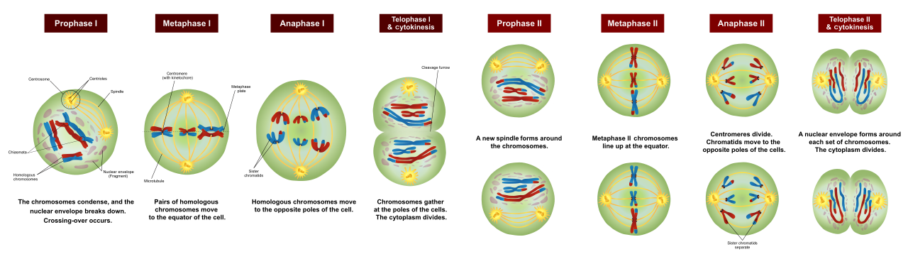
Meiosis I—Complications of prophase I (five subphases)
Prophase-I of meiosis is very long and complex, which is divided into five stages:
- Leptotene – Chromosomes gradually become clear.
- Zygotene—Synapsis of homologous chromosomes begins, forming a complex structure called the synaptonemal complex. This pair is called a bivalent or tetrad.
- Pachytene—The most important stage. Here, gene exchange (crossing over) occurs between the non-sister chromatids of homologous chromosomes—a process driven by the enzyme recombinase. This crossing over is the greatest source of variation in offspring.
- Diplotene – Disintegration of the synaptonemal complex and appearance of 'X' shaped structures called 'chiasmata'.
- Diakinesis – End of chiasmata and complete organization of spindle fibers.
Meiosis II
This is essentially the same as mitosis, which converts the haploid cells obtained after meiosis I into four haploid daughter cells. Thus, one diploid (2n) parent cell ultimately produces four haploid (n) gametes.
📐 Change of DNA amount in meiosis — formula/process: Genital-maternal cell (2n) → DNA duplication after S-phase (equivalent to 4n) At the end of meiosis I, at each pole → n (chromosome number halved) Four daughter cells at the end of meiosis II → n chromosomes each
🖼️ The main stages of meiosis—from one diploid cell to four haploid gametes
Homozygous versus meiotic division
| Number | Mitosis | Meiosis |
|---|---|---|
| 1 | the cell divides once | the cell divides twice |
| 2 | occurs in somatic cells | occurs in germ cells |
| 3 | found in both asexual and sexual reproduction | occurs only in sexual reproduction |
| 4 | DNA duplication occurs once during interphase | DNA duplication occurs only in the first interphase, not in the second. |
| 5 | The prophase is short and simple | Prophase I is the longest and most complex stage. |
| 6 | Crossing over does not occur, Chiasma is not formed | Crossing over occurs, chiasma is formed |
| 7 | The daughter cells have the same chromosome number as the parent (2n). | Half the chromosome number in daughter cells (n) |
During metaphase I, the maternal and paternal chromosomes randomly separate to either pole, and during pachytene, crossing over occurs—these two factors cause each offspring (even siblings) to look slightly different from their parents and from each other. This is the foundation of biological variation and evolution.
◆ Meiosis ◆ reductionist division ◆ synaptonemal complex ◆ bivalent ◆ crossing over ◆ Kazemeta ◆ gamete
15 Differences between plant and animal cells
So far, we've covered each cell organelle in detail individually. Now, putting these points together, the overall difference between plant and animal cells becomes very clear—a topic that's crucial for comparison questions in the RAS/IAS exam.
🖼️Comparative structure of plant cell and animal cell
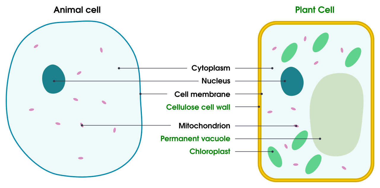
| Structure | plant cell | animal cell |
|---|---|---|
| cell wall | present (made of cellulose) | absent |
| cell size | Usually large and of a definite shape | relatively small and irregularly shaped |
| Vacuole | A large central vacuole is found | Vacuoles are small or absent |
| nutrition | Autotrophs – make their own food through photosynthesis (exception of fungi) | Heterotrophs – Photosynthesis does not occur (exception Euglena) |
| position of the nucleus | usually towards the edge of the cell | usually in the middle of the cell |
| Chloroplast | Present | absent |
| Lysosomes | very few or rare | usually present in abundance |
| Centriole | Usually absent (in some algae and fungi) | Present |
| Tarku fiber formation | are made from proteins present in the cytoplasm | are formed from centrioles |
| Plasmodesmata | Present | absent |
| Spherosome | Present | absent |
| stored food | as starch | in the form of glycogen and fat |
| cytoplasmic division | from the center to the edges by the cell plate | grooves from the edges towards the center |
Remember the plant cell as a 'house' with its own strong walls (cell wall), a large water storage room (central vacuole), and a solar panel-like roof (chloroplast)—this house can make its own food (autotrophic). Remember the animal cell as a 'flat' with no walls, no solar panel—so it has to get its food from the market (heterotrophic).
◆ plant cell ◆ animal cell ◆ Self-sufficient ◆ Heterotroph ◆ Central vacuole
16 Major discoveries of cytology
Let us now look at all the scientists and their discoveries that we read about in this entire chapter, in a systematic manner, in one place – this table will prove to be very helpful in quick revision of the entire chapter.
| Scientist (Year) | Discovery/Contribution |
|---|---|
| Robert Hooke (1665) | First observed cell (cell wall) in cork, described in 'Micrographia' |
| Antonie van Leeuwenhoek (1674) | First observed living cell |
| Robert Brown (1831) | Discovery of the Nucleus |
| Purkinje (1840) | named protoplasm |
| von Moll (1846) | introduced the importance of protoplasm |
| Rudolf Virchow (1855/1858) | 'New cells are formed by the division of preexisting cells' |
| Haeckel (1865) | Use of the term 'plastid' |
| Mischer (1868) | discovery of nucleic acids |
| W. Fleming (1879/1882) | Mitosis and the use of the term 'chromatin'; mitochondria were named 'phyla' |
| Strasburger (1879) | Difference between cytoplasm and nucleoplasm, cell division in plants |
| Schimper (1883) | Study of chloroplasts |
| Altmann (1886/1894) | Discovery of mitochondria, named 'bioplast' |
| Waldeyer (1888) | named chromosome |
| T. Baveri (1888) | named the centrosome |
| C. Benda (1897) | named 'mitochondria' |
| Camillo Golgi (1898) | discovery of the Golgi apparatus |
| Sutton and Baverie (1902) | proposed the chromosome theory |
| Farmer and Moore (1905) | Description of Meiosis |
| Noll and Ruska (1932) | invention of the electron microscope |
| Claude (1941) | Ribosomes were first observed |
| Keith Porter (1945/1953) | named the endoplasmic reticulum (ER) |
| D. Dewey (1955) | Discovery of lysosomes |
| Singer and Nicholson (1972) | Introduced the Fluid Mosaic Model |
◆ cell Biology ◆ Micrographia ◆ Fluid mosaic model ◆ electron microscope
• The cell is the smallest structural and functional unit of a living organism; it was discovered by Robert Hooke (1665) and Schleiden-Schwann (1838–39) formulated the cell theory, to which Virchow (1855) added 'Omnis Cellula e Cellula'. • Viruses are an exception to the cell theory because they do not have the ability to divide independently and do not have complete cellular components. • Based on the organization of the nucleus, cells are of two types – prokaryotic (without a well-organized nucleus, like bacteria) and eukaryotic (with a well-organized nucleus, like plants and animals). • The cell wall (only in plants, fungi, and bacteria) gives shape and strength to the cell, while the cell membrane (in all cells) is a selective membrane composed of a phospholipid bilayer and proteins, according to the fluid mosaic model, which regulates the movement of substances by passive and active transport. • Mitochondria are the cell's 'powerhouse' where ATP is produced through aerobic respiration; chloroplasts/plastids carry out photosynthesis—both are semi-autonomous organelles. • Endoplasmic reticulum (protein/fat synthesis), Golgi bodies (packaging-secretion), ribosomes (protein production), centrosomes (spindle fiber production), lysosomes ('suicide bags' - intracellular digestion) - all these organelles together run the cell like a well-organized 'factory'. • The nucleus is the control center of the cell, in which chromatin material condenses into chromosomes during cell division—humans have 46 (23 pairs) chromosomes. • Mitosis promotes growth and repair, and the daughter cells retain the same chromosome number as the parent cell; while meiosis produces gametes for sexual reproduction, where the chromosome number is halved and genetic diversity is created through crossing over. • Plant cells have cell wall, large vacuole, chloroplast and autotrophic nutrition, whereas animal cells lack these and are heterotrophic. • This entire journey of cell biology—from Hooke's cork to Singer-Nicholson's fluid mosaic model—shows that even the greatest secrets of life are hidden within the smallest unit—the cell.